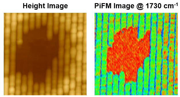
e-Beam Damage on ArF Photo Resist
This slide shows e-beam damage on ArF photoresist.
The AFM topography image reveals surface modifications caused by e-beam exposure.
The PiFM image highlights compositional changes in the exposed regions, clearly distinguishing them from the unaffected areas.
Scanning Conditions
- System: AFM-IR
- Sample: Sample: PR pattern sample
- Scan Mode: PiFM (Channel: Z Height, PiFM Amplitude)
- Scan Rate: 1 Hz
- Scan Size: 1 µm × 1 µm
- Pixel Size: 512 × 256 pixels
Related Contents

System Webinars
Deeper insights into 2D-Materials and related Microcrystals by Imaging Spectroscopic Ellipsometry (ISE) and Imaging Mueller Matrix Ellipsometry (IMME)

Application Talks
Imaging Müller Matrix Ellipsometry for Quantifying Dielectric Tensors of Molecular Microcrystals as well as Analyzing Engineered Microstructures

Application Talks
Giant Optical Anisotropy and High Refractive Index in van der Waals Materials
×


