
Origami DNA
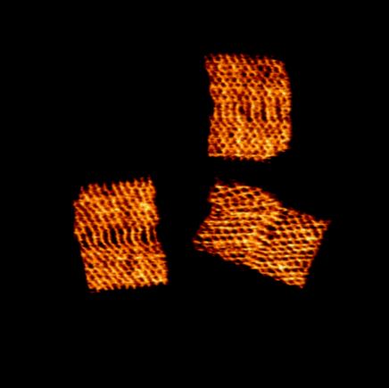
Origami DNA was imaged in liquid using Tapping mode to obtain surface topography. The scan image shows rectangular DNA origami structures with internal patterns, with height variations up to 2.6 nm.
Scanning Conditions
- System: NX10
- Scan Mode: Tapping in liquid
- Scan Rate: 2 Hz
- Scan Size: 300 nm × 300 nm
- Pixel Size: 512 × 512 pixels
- Peak-to-valley: 2.6 nm
• Sample courtesy: PhD. Luca Piantanida from Micron School of Materials and Engineering at Boise State University, USA
Related Contents
.jpeg)

×