
Lung cancer cell
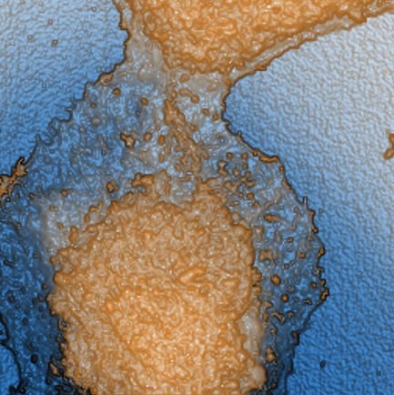
A lung cancer cell was imaged using Scanning Ion Conductance Microscopy (SICM). The topographical image reveals detailed surface structures of the cell, showing a peak-to-valley height variation of approximately 8.17 µm.
Scanning Conditions
- System: NX12
- Scan Mode: Scanning Ion Conductance Microscopy
- Scan Rate: 0.3 Hz
- Scan Size: 60 µm × 60 µm
- Pixel Size: 256 × 256 pixels
- Peak-to-valley: 8.17 µm
Related Contents
.jpeg)

×