
Giardia in liquid
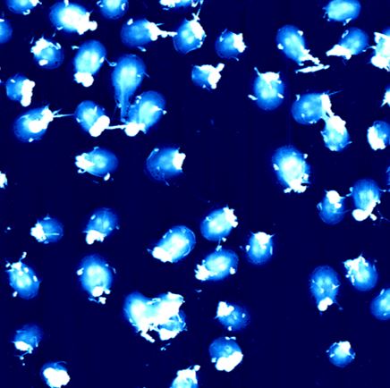
A Giardia sample in liquid was imaged using Tapping mode. The topographical image clearly shows the surface structure of the microorganism, with a peak-to-valley height variation of approximately 2.8 µm.
Scanning Conditions
- System: NX12
- Scan Mode: Tapping mode
- Scan Rate: 0.2 Hz
- Scan Size: 100 µm × 100 µm
- Pixel Size: 512 × 512 pixels
- Peak-to-valley: 2.8 µm
Related Contents
.jpeg)

×