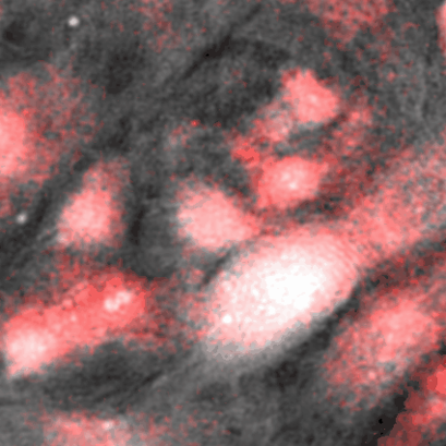
Speaker : Dr. Bartlomiej Zapotoczny Affiliation : Polish Academy of Sciences, Poland Region : Europe Year : 2022 Language : English Liver sinusoidal endothelial cells (LSECs), due to their unique morphology, are an excellent research target for developing high-resolution imaging methods [1]. They possess transcellular pores called fenestrations. Fenestrations in LSECs have diameters in the range of 50-350 nm [2], being largely beyond the resolution limits of classical optical microscopy, limited by light diffraction. Fenestrations in LSECs play a crucial role in realizing the transport of substances, especially lipoproteins, between blood and hepatocytes in the liver. Reduced LSEC porosity has been shown to be associated with chronic liver diseases such as hepatitis, steatosis and cirrhosis. Furthermore, the number of fenestrations decreases with age. In vitro studies have shown that LSEC porosity can be altered pharmacologically [2,3]. Due to the great importance of fenestrations diameters, acting as a functional sieve for particles of a certain size into and out of the liver, the determination of the "true" fenestration size is extremely desirable. Results obtained in recent years using a wide range of micro- and nanoscopic methods applied to LSECs in order to assess drug and toxin effects will be presented. All techniques have their limitations, making it difficult to accurately determine the size of fenestration. A quantitative and qualitative description of the differences will be made in a correlative manner. Scanning electron microscopy (SEM) was combined with optical nanoscopy methods (STED, SIM) (Fig.1). Additionally, atomic force microscopy (AFM) was used in combination with SEM and STED, all in order to better understand the differences between literature reports on fenestration dimensions. Research supported by the National Science Centre under the project "SYMFONIA 3", (UMO-2015/16/W/NZ4/00070), SONATA 15", (UMO-2019/35/D/NZ3/01804), and Research Council of Norway Nano2021 (288565). [1] Szafranska K.,[...], Zapotoczny B. Nanophotonics, 2022. doi. 10.1515/nanoph-2021-0818; [2] Zapotoczny B. et al., Hepatology, 2019 doi. 10.1002/hep.30232; [3] Zapotoczny B. et al., Biophysical Reviews, 2020 doi. 10.1007/s12551-020-00699-0;