


Melamine cyanurate


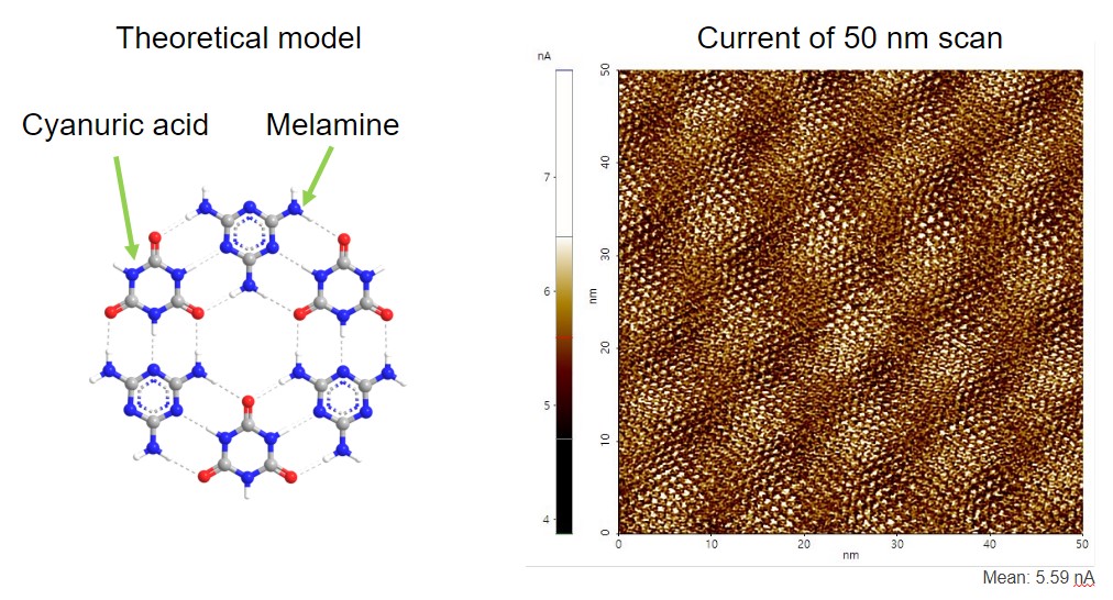
A conductive AFM scan of melamine-cyanuric acid network on HOPG acquired with internal C-AFM on NX10. This is a remarkable image, since previously only STM was able to achieve such resolution in electrical modes, shows molecular structures of cyanuric acid and melamine.
Scanning Conditions
- System: NX10
- Sample Bias: 0.17 V
- Scan Mode: Conductive AFM (C-AFM)
- Scan Rate: 20 Hz
- Scan Size: 50 nm×50 nm
- Pixel Size: 512×512
- Cantilever: NSC36/Pt C type (k= 0.6 N/m, f= 65 kHz)
×