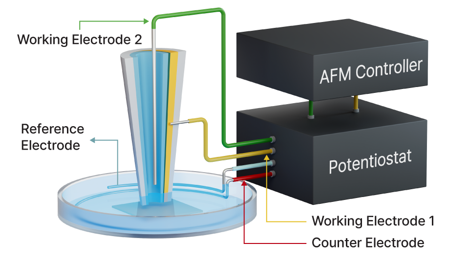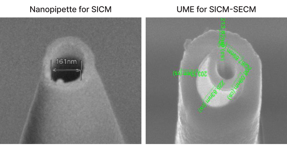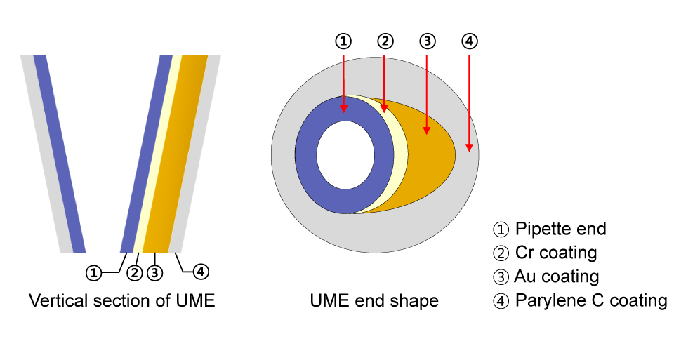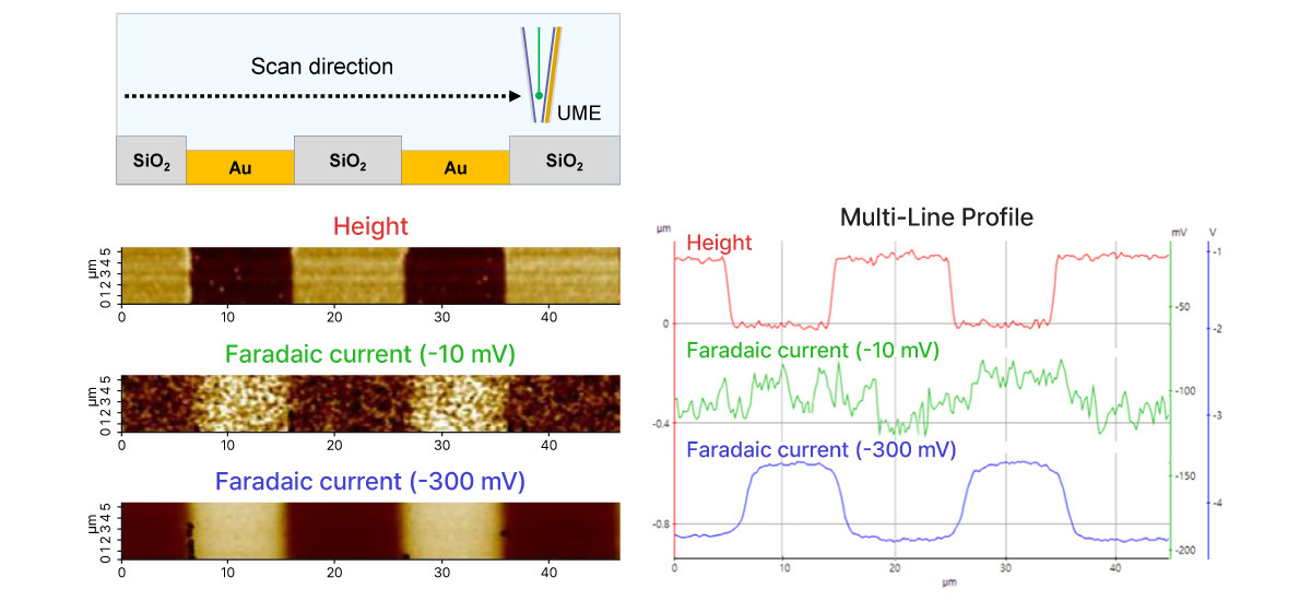
Scanning Ion Conductance Microscopy-Scanning Electrochemical Microscopy
SICM-SECM
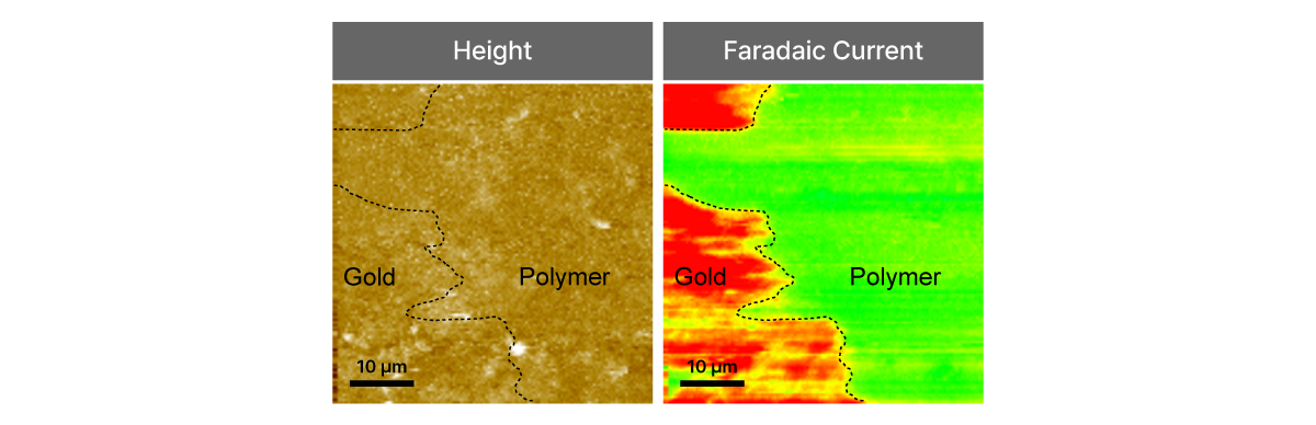
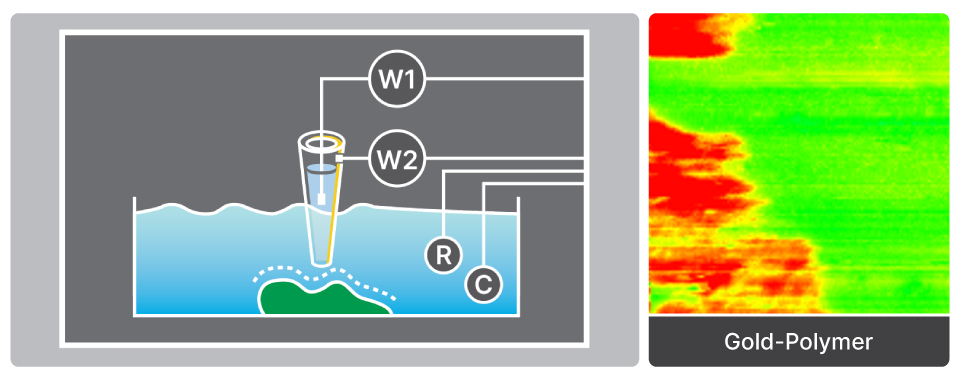
Combines SICM with SECM for simultaneous surface topography and electrochemical activity imaging