Scanning Electrochemical Cell Microscopy
SECCM
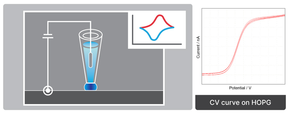
Localized electrochemical characterization employing a nanopipette as an electrochemical cell to map chemical reactivity
What is SECCM
(Scanning Electrochemical Cell Microscopy)
SECCM measures localized electrochemical activity by employing a nanopipette to create a confined electrochemical cell at the sample surface, enabling high-resolution mapping of redox reactions, catalyst behavior, and surface modifications under ambient conditions.
SECCM employs a nanopipette filled with electrolyte and equipped with an internal pipette electrode. The nanoscale droplet at the pipette tip forms a confined electrochemical cell when it contacts the substrate, isolating the region of interest for localized measurement.
An electrode is inserted inside the nanopipette to collect the electrochemical signal. Approach control is achieved by monitoring the ionic current: when the meniscus contacts the surface, a sharp surge in current halts the approach. The system detects this signal and immediately stops the Z scanner movement, maintaining precise tip-sample spacing. The electrochemical species in the confined meniscus undergo an electrochemical reaction when a bias is applied between the nano-pipette electrode and the working electrode placed on the XY scanner.
Reasons to Use This Mode
Unlike other pipette-based SPM techniques such as SICM or SICM-SECM, SECCM utilizes an electrolyte-filled nano-pipette to form meniscus contact with a sample surface to create an electrochemical cell at the end of the nano-pipette so that the measurement in ambient conditions is possible. That is, only a very local area is made into a liquid condition and the electrochemical reaction occurring in that area is observed. Also, the response area (electrochemical cell) can be controlled by the inner diameter size of the nano-pipette.
Applications and Use Cases
AFM and SECCM measurements on HOPG/ITO substrate to determine surface morphologies and to map EC signal contrasts of these two different materials. The regular AFM and the SECCM measurements are conducted on the same area. Using the confined meniscus contact on HOPG surface, as the working electrode, nano-scale imaging could even distinguish the EC signal of the graphene basal plane, multi-layered graphene, and graphene with a conductive substrate. Different layers and step edges are visible in the height image that well align with distinctive features in the SECCM mapping image visualizing different EC activities of different graphene layers and step edges.
  • Sample: HOPG Multilayer on ITO
  • System: NX10
  • Scan Size: 50 µm × 50 µm