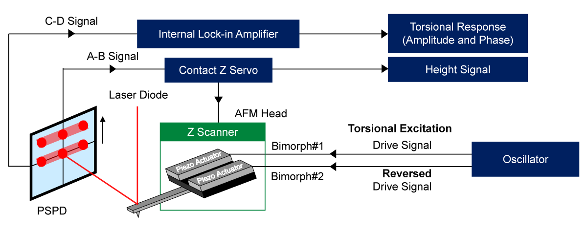
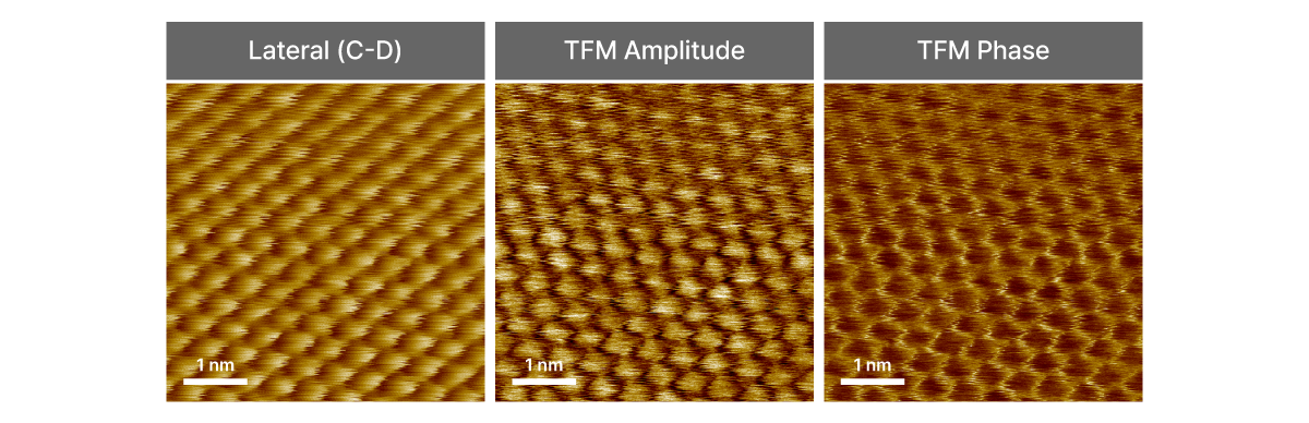
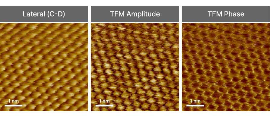
Torsional Force Microscopy
TFM
Lateral force mapping by detecting torsional deflections of the cantilever during scanning over the sample surface