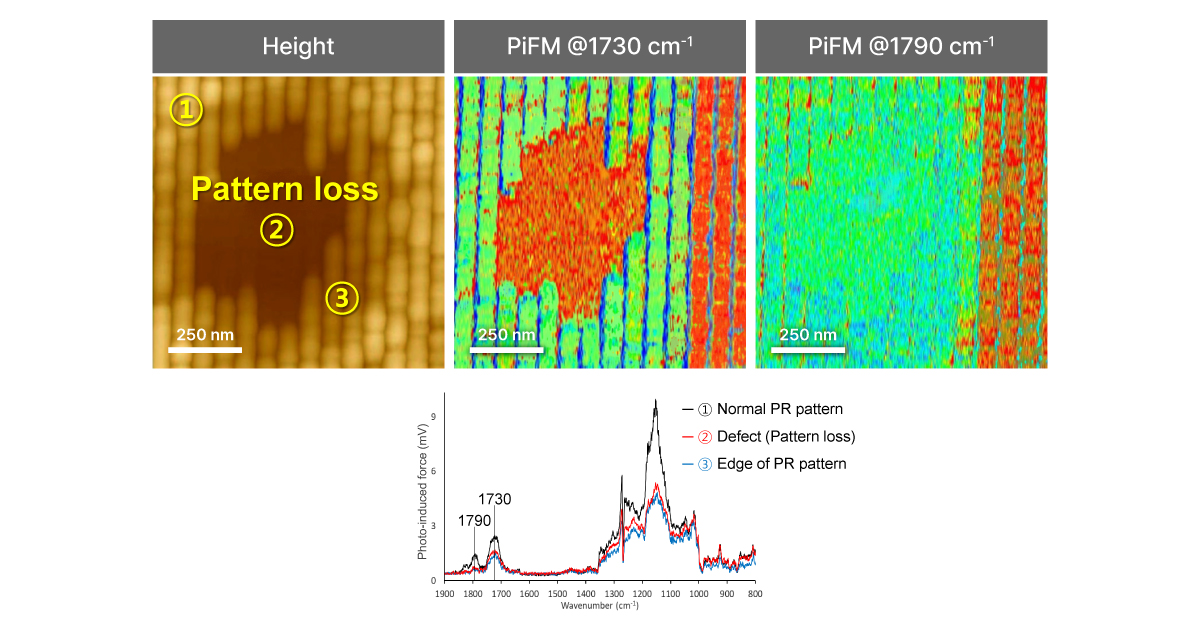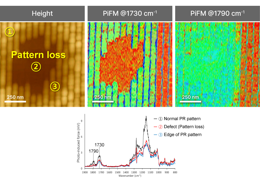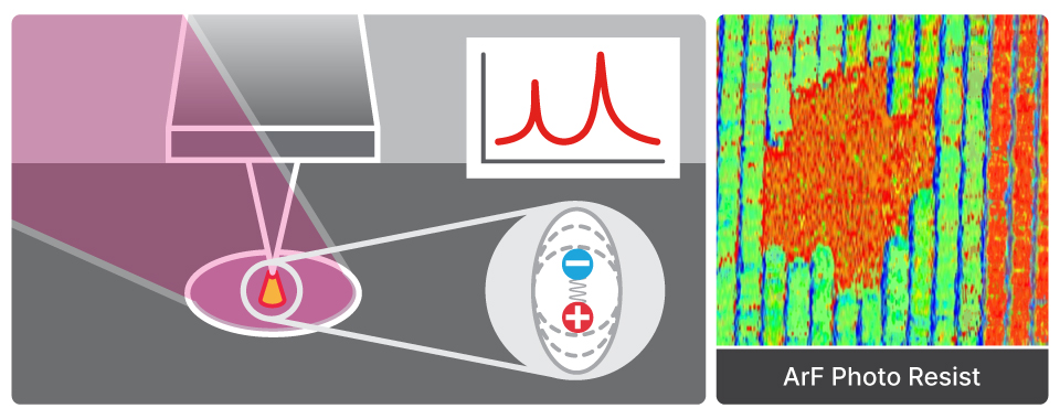
Photo-induced Force Microscopy
PiFM
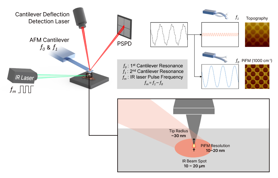
Chemical and compositional nano-imaging by detecting photothermal-induced tip–sample forces at specific infrared absorption frequencies