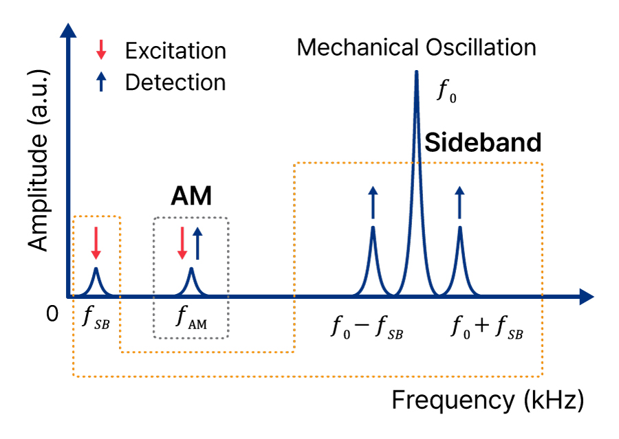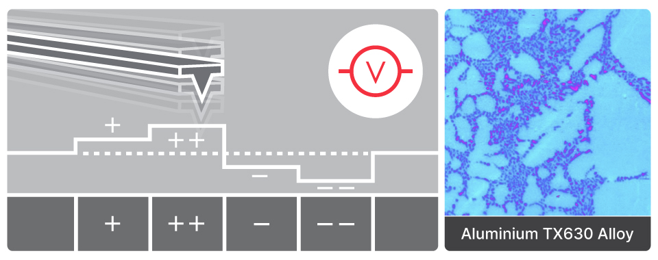
Sideband Kelvin Probe Force Microscopy
Sideband KPFM
Enhanced surface potential sensitivity and spatial resolution through resonance frequency detection