


Application Options
Live Cell Study with Live Cell Chamber (SICM & AFM)
The live cell chamber creates an ideal environment for cells, improving their life expectancy during long measurement durations through controlled temperature, pH, and humidity at optimal conditions. Experiments with the live cell chamber have demonstrated cell survivability of more than 20 hours.
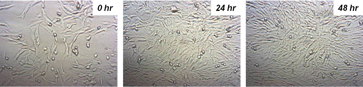
Human fibroblast cells in the Live Cell Chamber of NX-Bio survive over 48 hours.
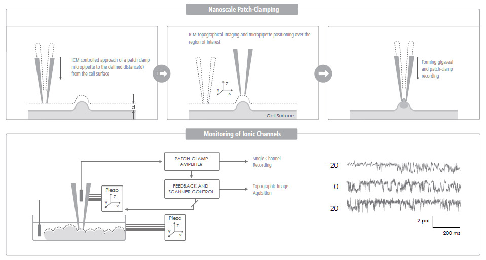
Ion Channel Recording of Targeted Patch Clamping
Conventional Patch Clamping is an optical microscope view-based technique used to monitor a single living cell's ion channel activity—a key quantifier of various cellular activities. Targeted Patch Clamping is the SICM-based version of this technique that enables the detection of ion channel activities of specific subcellular structures.
SICM + Patch Clamping = Targeted Patch Clamping
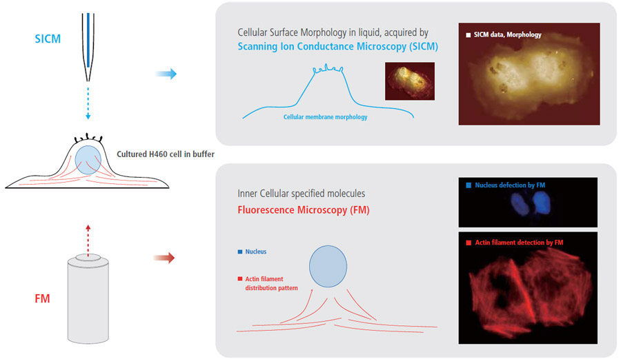
More Comprehensive Cell Biology Study, by Integrating Fluorescence Microcopy with Park SICM
Combining fluorescence microscopy (FM) techniques with Park SICM can create new benefits and provide comprehensive information for cell biology studies that cannot be obtained when using only one of those techniques. While monitoring external cellular surface morphology with SICM, the internal cellular behavior can be observed by FM.
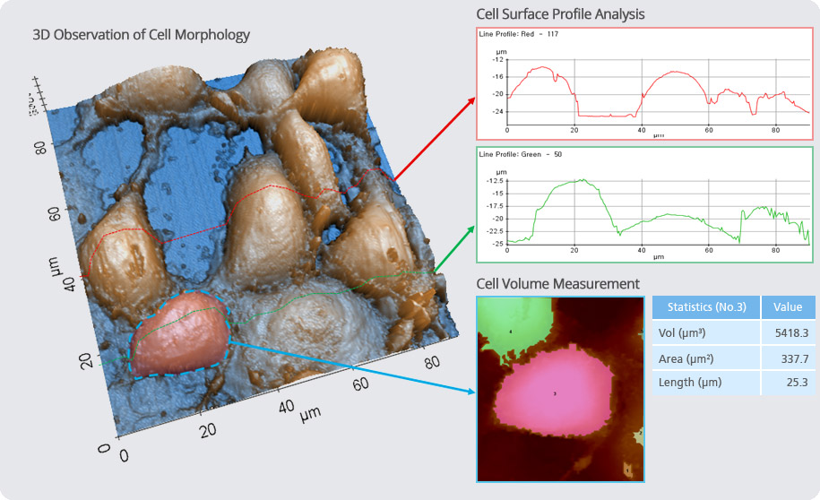
Structural & Physical Property Characterization of Cell in XEI
Software Option
SmartScanTM – Data Acquisition
SmartScanTM is a data acquisition software that provides all user controls of Park AFM measurements. The friendly user-oriented interface of SmartScanTM provides easy operation of the AFM.
• Simultaneous data acquisition of up to 16 images
• Maximum 4096 × 4096 pixels image size
• Dedicated Force-distance and I-V spectroscopy with batch processing
• Cantilever spring constant calibration
XEI – Image Processing and Analysis
XEI is the AFM image processing and analysis program. Its powerful processing algorithms make analyses easy and streamlined. With its most advanced and versatile imaging features, AFM users can obtain essential and critical information from their experiment.
• Image analysis of line profile, region, 3D rendering
• Spectroscopy data analysis module (F-d, I-V)
• Directly copy/paste to presentation program
• Multiple image comparison
• Image overlay of two different images
Image Overlay: SICM Topography + Fluorescence Microscopic Image
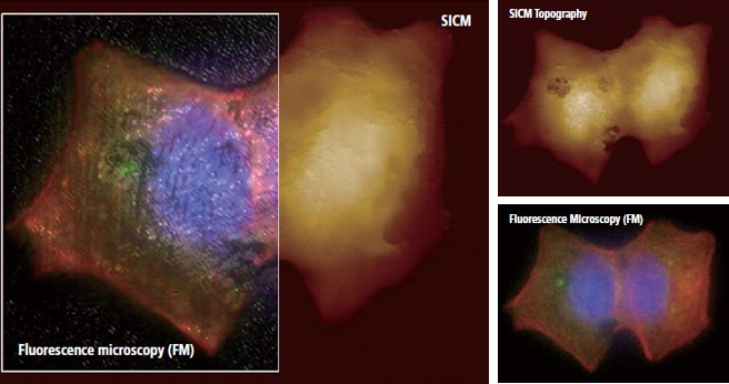
Park Systems’ Image overlay software allows to combine fluorescence microscopic image onto SICM topography accurately. This dedicated software helps a complicate combining process much easier .


