- SICM Technology
- AFM Technology
- SICM + AFM
The SICM of Park NX12 is the next generation nanoscale microscope for life science
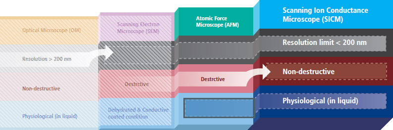
Park SICM can acquire biological images at nanoscale in physiological conditions, attaining high resolution of less than 200 nm. The biological images obtained from SICM are free from morphological deformation, which can occur from scanning electron microscopy (SEM) or even AFM systems.
Park SICM uses nanopipettes
In Scanning Ion Conductance Microscopy developed by Park Systems (Park SICM), a glass nanopipette filled with an electrolyte acts as an ion sensor that provides feedback on its location relative to a sample completely immersed in liquid. The pipette tip maintains its distance from the sample by keeping the ionic current constant. In comparison, AFM typically relies on interaction of forces between its probe tip and the sample.
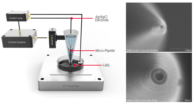
AFM uses a micro-thin cantilever and tip as a probe. For Park SICM, the pipette is a probe with an inner diameter ranging from 80 to 100 nanometers for pipettes made of glass and 30-50 nanometers for those made of quartz.
No Force, Non-Contact Imaging in Liquid
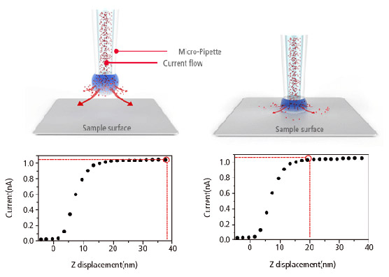
Similar to Scanning Tunneling Microscopy (STM) operating in ambient air, the Park SICM operates in liquid without making physical contact with the sample. Electrodes on either side of the sample and pipette produce ionic current that flows through the surrounding solution. A sensor measures the current flow, which decreases as the distance between the pipette and sample becomes smaller, and monitors the distance between the pipette and the sample to obtain the topology.
Park SICM Can Image All Cell Types
- Park SICM can image even the softest cells such as the neuron cells, live—something that’s impossible with any other microscopy techniques.
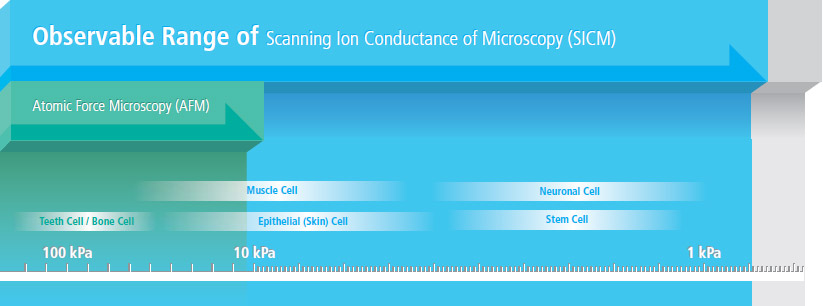
- SICM can even image suspended network of neurons
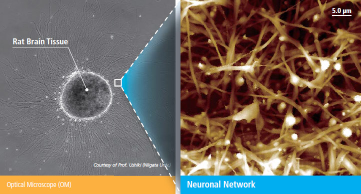 Courtesy of Prof. Ushiki (Niigata Univ., Japan)
Advanced Park AFM Technology Enables Accurate Force-distance Spectroscopy
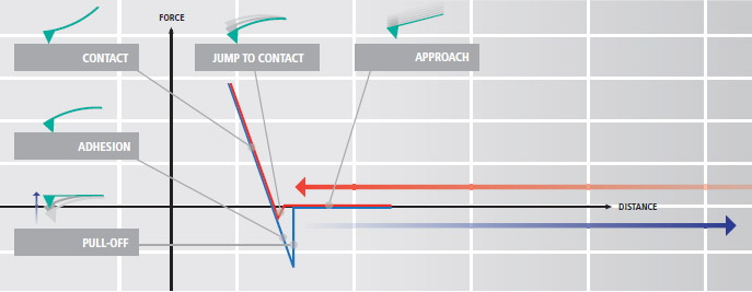
Force-distance (FD) spectroscopy using an AFM is a beneficial tool to characterize bio-mechanical properties of various biological materials. In FD spectroscopy, the cantilever tip touches the sample surface with a user prescribed amount of force accurately applied using the AFM’s Z scanner. Park AFM’s industry leading low noise Z detector allows the researcher to control Z scanner movement to apply an exact amount of force very accurately to a sample surface during the FD spectroscopy. This enables the researcher to collect detailed bio-mechanical characterization data at the nano-newton scale.
- Nanomechanics of Single Muscle Fibers by AFM
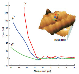 International Nano-Conference(ICN+T), Basel (CH), 2006, Noemi Rozlosnik Technical University of Denmark
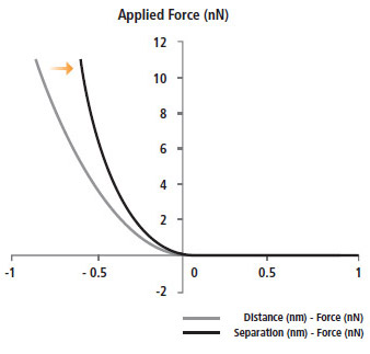
- Force Distance Spectroscopy measures the mechanical interaction force between the tip end and the sample.
- The force-distance curve is acquired by indenting the cantilever to sample surface.
Advanced Bio-mechanical Property Measurement by Calculating Elastic Modulus (Young’s Modulus)
The Herzian and Oliver Pharr models are calculated automatically from the Park AFM's accurate FD spectroscopy data to determine the elastic modulus (Young's modulus). Both of these calculation methods are included in Park XEI, the data analysis software in Park NX12. They strengthen the biomechanical data verification of FD curves obtained in your experiments.
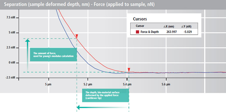
- Acquiring the actual depth, sample deformed by applied force
(separation - force curve)
- Calculating Young's Modulus in Hertzian model
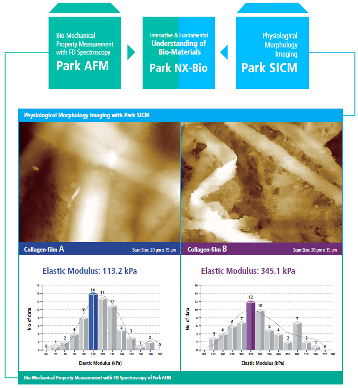
Park SICM and Park AFM Technologies Put Together
Outstanding Investigation Tool for Biological Research by Combining Physiological Morphology with Bio-Mechanical Property Measurements
Park NX12 combines Park SICM’s ability to interpret morphology under true physiological conditions and Park AFM’s capacity to acquire bio-mechanical property data (elastic modulus) accurately. This enables researchers to understand the fundamentals of their biological materials at a deeper level.