


Twisted Bilayer Graphene - 2


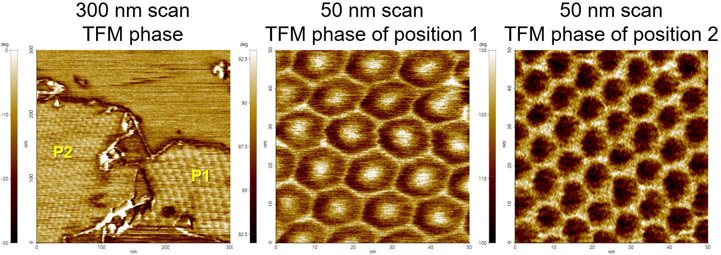
Twisted bilayer graphene (tBG) stacked on hBN forms a nanoscale moiré superlattice, where local stacking configurations create region-dependent structural and electronic variations.
TFM phase imaging clearly visualizes the moiré superlattice and reveals distinct contrast and periodicity differences between position 1 and position 2, enabling high-resolution mapping of local stacking and heterostructure domains.
Scan Conditions
- System: NX10
- Scan Mode: TFM
- Scan Size: 300 nm×300 nm, 50 nm×50 nm
- Scan Rate: 16 Hz, 8 Hz, 10 Hz
- Pixels: 512×512
- Cantilever: SCOUT 70
(k=2 N/m, f=70 kHz)
*Sample courtesy: Prof. Tomoki Machida, Yuta Seo, Institute of Industrial Science, University of Tokyo, Japan
×