


Teflon Defect


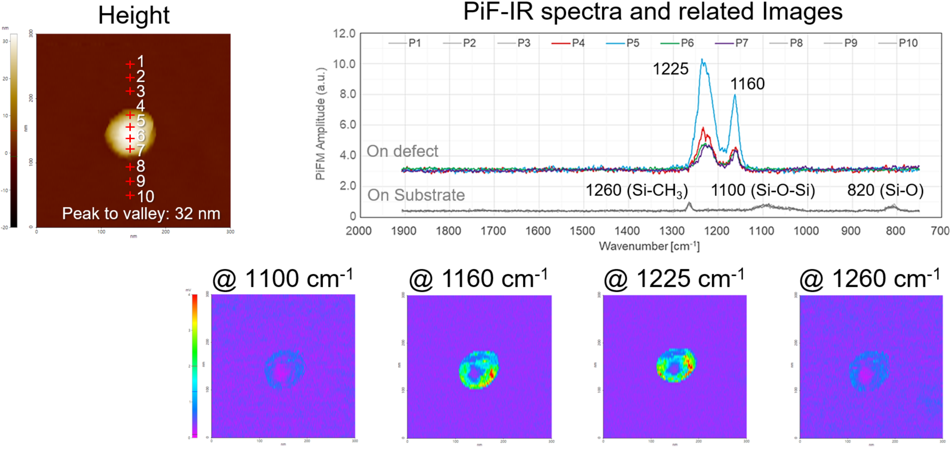
PiFM enables defect analysis beyond simple morphology by acquiring local IR spectra to identify the chemical composition of the Teflon defect and by imaging at selected wavenumbers to map the full defect distribution.
Scan Conditions
- System: FX200 IR
- Scan Mode: PiFM
- Scan Size: 300 nm×300 nm
- Scan Rate: 0.1 Hz
- Pixels: 512×64
- Cantilever: PPP-NCHPt
(k=42 N/m, f=330 kHz)
×