Park SICM
Park Scanning Ion Conductance Microscopy
In Scanning Ion Conductance Microscopy developed by Park Systems (Park SICM), a glass nanopipette (a pipette in the nanoscale) filled with an electrolyte, acts as an ion sensor that provides feedback on its location relative to a sample completely immersed in a liquid. The pipette-tip maintains its distance from the sample by keeping the ionic current constant. In comparison, Atomic Force Microscopy (AFM) typically relies on interaction of forces between a probe tip and the sample.
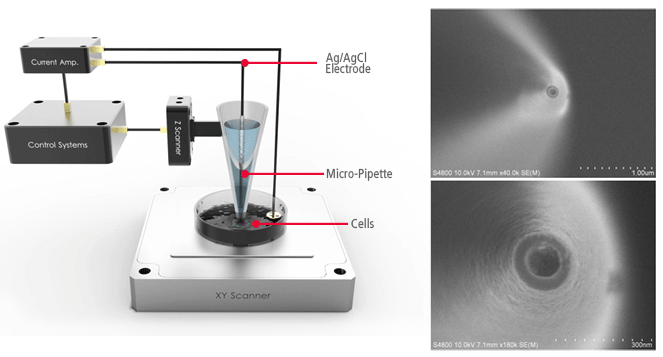
AFMs use a micro-thin cantilever and tip as a probe. For Park SICM, the pipette is a probe with an inner diameter ranging from 80 to 100 nanometers for pipettes made of glass and 30-50 nanometers for those made of quartz.
No Force, Non-Contact Imaging in Liquid
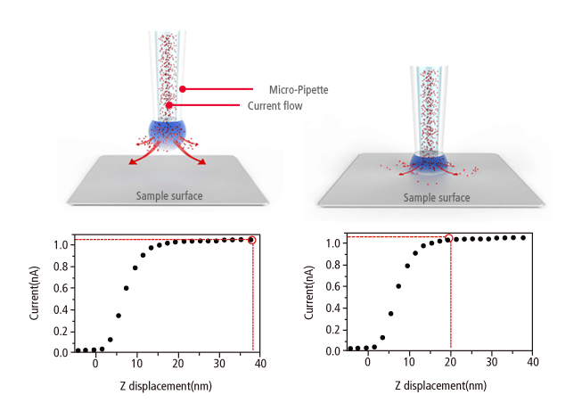
Similar to Scanning Tunneling Microscopy (STM) operating in ambient air, the Park SICM operates in a liquid without making physical contact with the sample. Electrodes on either side of the sample and pipette produce an ionic current that flows through the surrounding solution. A sensor measures the current flow, which decreases as the distance between the pipette and sample becomes smaller. This monitors the distance and directs the positioning of pipette and sample to maintain non-contact status.
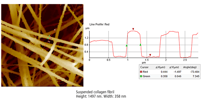
The Park SICM image on the right is suspended collagen fibril. The suspended multi-fibrils have a very complex structure, floating a several levels just microns from the bottom substrate in the liquid. This is an impossible configuration for traditional Atomic Force Microscopy as penetration into the liquid or the force of a physical probe displaces the suspended fibrils.
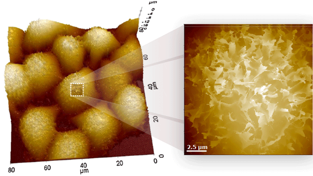
Live Cell Membrane Imaging with Park SICM
The cell membrane separates the interior of cells from the outside environment. Observing the functioning of living cell membranes is an increasingly important element of cell research, but it is very difficult to monitor a live cell membrane at the nanometer scale. The transparency of the membrane makes it nearly impossible to observe with optical microscopy and the membrane is too fragile to endure the force involved with AFM or Scanning Electron Microscope probes.
Park SICM, in contrast, applies no force to the sample surface and is ideal for nanoscale imaging of soft live cell membranes. The Park SICM image on the left shows live Hela cells. The cells remained live and stable throughout the duration of Park SICM imaging, showing no signs of physical deterioration. The structural complexity of live cell membranes like this is almost impossible to observe with optical microscopy or AFM