Thursday, 9 April 2020
- 10:00 am – 11:30 am
(GMT)
London, Dublin - 11:00 am – 12:30 pm
(CEST)
Berlin, Paris, Rome - 18:00pm – 19:30 pm
[UTC+9]
Seoul, Tokyo -

Abstract
Kelvin Probe Force Microscopy (KPFM) is a scanning probe method that utilizes electrostatic forces between a nanoscopic, conductive tip and a sample for material characterization by mapping the distribution of the surface potential [1,2].
In the standard lift mode KPFM [3], the sample is first scanned in dynamic mode at close distance in order to retrieve its topography. During the second pass, the tip is retracted at a given lift distance and traces back the topography while electrically excited at the lever eigenmode frequency. Since the conductive tip is sensitive to the local gradient of the electrical field, one can minimize the amplitude or phase shift of the probe caused by such a gradient by applying a counteracting voltage on the tip, which would exactly compensate the sample potential at each pixel. This allows to detect and image the potential variation along each line of the scanned frame.
However, this dual-pass mode has several technical drawbacks. First, due to the lift height the electrical signal measured by the tip is necessarily an averaged contribution of a larger area of the sample during second pass, which implies a loss in lateral resolution. Second, since the tip needs to scan each line twice, a longer image acquisition time must be taken into account. On the other hand, a simultaneous mechanical and electrical excitation of the lever at its resonant frequency would cause a severe crosstalk between topographic and electrical signals.
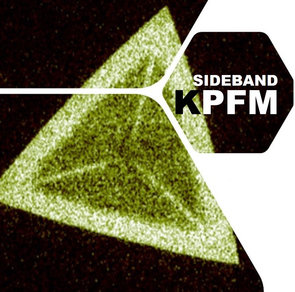
The new sideband KPFM mode allows for simultaneous imaging of the topography and surface potential with a high spatial potential resolution and improved sensitivity with respect to lift mode and other KPFM (e.g. off-resonance) methods. Here, the topography is detected at the resonance of the cantilever, while the electrical drive of the KPFM is applied at much lower frequencies between 1 and 4 kHz. Due to frequency mixing, satellite peaks or sidebands appear in 1 and 4 kHz from the resonance. The parallel detection of both excitations is feasible due to the presence of multiple lock-ins in the Park NX default electronics. Since the KPFM signal is detected on the sidebands and the topography is detected on the resonance, both signals can be measured simultaneously.
This webinar will provide a theoretical introduction to KPFM and show live examples of sideband KPFM experiments using the Park NX20 Large Sample AFM.
[1] U. Zerweck et al., Phys. Rev. B 71 (2005) 125424.
[2] A. Axt et al., Beilstein J. Nanotechnol. 9 (2018) 1809-1819.
[3] H. O. Jacobs et al., Ultramicroscopy 69 (1997) 39-49.

Presented By :
Dr. Andrea Cerreta - Application Scientist Park Systems Europe, Mannheim, Germany
acerreta@parksystems.com
Dr. Andrea Cerreta is an Application Scientists at Park Systems Europe, where he focuses on application development and support for the academic sector. He received his Ph.D. in Physics from the Ecole Polytechnique Fédérale de Lausanne (EPFL), Switzerland. He did his further doctoral work at the Solid State Physics Group of Université de Fribourg, Switzerland, which focused on studying electrical and magnetic properties of organic spin valves and spin polarized currents in superconducting materials, grown by means of Pulsed Laser Deposition, and characterizing the DC and AC transport properties of magnetic and superconducting samples. His expertise also spans the Frequency Modulation Atomic Force Microscopy in UHV for the study of biomolecules.


