


Brian Choi, Bio-application scientist
For more information, please contact app@parksystems.com
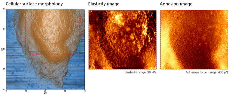
To study cell at fundamental level, cell’s physical properties (morphology and stiffness/elasticity/adhesion force) must be measured accurately.

- Analyze cell at fundamental level, by acquiring cell morphology and quantitative mechanical property image of cell simultaneously
- PinPoint™ mode allows users to visualize elasticity (Young’s modulus) and adhesion force as an image providing quantitatively calculated values
PinPoint mode™ is a newly developed AFM imaging mode that acquires topographical data and the FD data at each pixel simultaneously. Using PinPoint™ mode, cell morphology and the quantitative nanomechanical properties of cell can be obtained at the same time. The height information constituting the morphology of the sample surface is determined when the designated stiffness threshold is reached. The elasticity (stiffness) at certain positions of the sample surface can be obtained the same way as in the FD spectroscopy. The stiffness can be obtained from the slope of FD curve from contact point to deflection threshold generated during the PinPoint mode™ operation. Applying Hertz model, designed for soft and elastic samples, the user can calculate the elasticity (Young's modulus) from the slope, too. The adhesion image is generated based on the maximum adhesion force value.
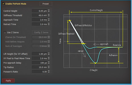 PinPoint™ mode Control
Cell Mechanical Property Study with PinPoint™ mode
Park Cell Analysis Systems
|
|
|
|
| Park NX12-Bio | Park NX10 | Park XE7 | |
| Scanning Ion Conductance Microscopy (SICM) | |||
| Atomic Force Microscopy (AFM) with liquid probe hand | |||
| Inverted Optical Microscopy (IOM) | |||
| Live Cell Chamber |