


Brian Choi, Bio-application scientist
For more information, please contact app@parksystems.com
Reference: Bing-Chen L., Douglas C. E., Heping M. (2013) SICM a nanotechnology for biological studies in live cells.
Frontiers in physiology. Vol 3. article 483
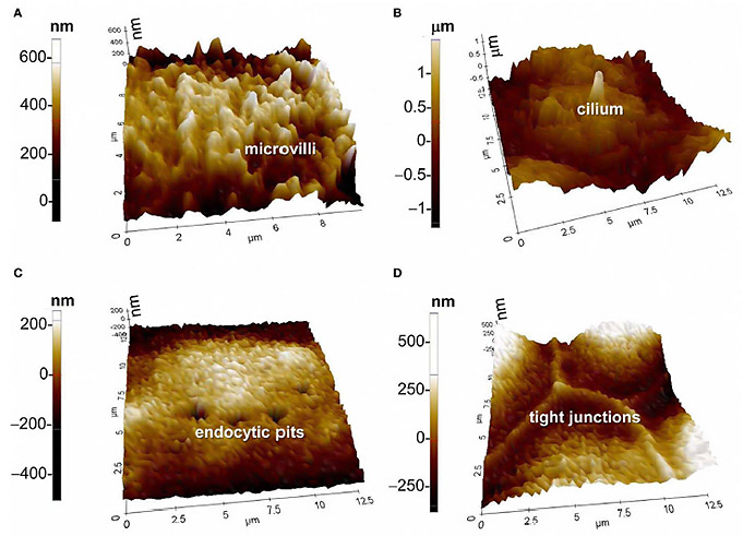
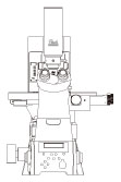
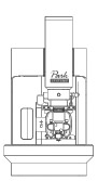
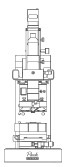
Scanning ion conductance microscopy (SICM) is a powerful tool to study and investigate cell function and important physiological processes related to specialized features on the cell membrane. SICM can visualize specialized structures such as microvilli, cilia, endocytic pits, and tight junctions of apical membranes of epithelial cells in physiological conditions. Conventionally, such delicate cell structures are observable only in fixed and dehydrated status using scanning electron microscopy and atomic force microscopy. However, to understand cell function and facilitate the study of important physiological processes, the direct observation of fine structures in living cell membranes is vital.
- Study specialized cell membrane structures such as microvilli, cilia, endocytic pits, and tight junction
- Investigate cell function and important physiological process, related with such specialized structures on cell membrane
SICM allows researchers to observe the fine structural and functional changes of membranes in living tissue. Investigating such structures with SICM reveals many new facts. For example, multiple microvilli can either form ridges or break into small isolated structures. In addition, the height of microvilli can be rearranged in response to cell volume changes. Also, the morphological changes of tight junctions occur after hypertonic stress. The role of tight junctions is known to maintain epithelial monolayer integrity. The images shown below, taken by a Park SICM, clearly describe such fine structures of microvilli, cilia, endocytic pits, and tight junctions in mouse cortical collecting duct mpkCCDc14 cells.
 Specialized Cell Membrane Structures
Park Cell Analysis Systems
|
|
|
|
| Park NX12-Bio | Park NX10 | Park XE7 | |
| Scanning Ion Conductance Microscopy (SICM) | |||
| Atomic Force Microscopy (AFM) with liquid probe hand | |||
| Inverted Optical Microscopy (IOM) | |||
| Live Cell Chamber |