


Alyssa Miller is currently enrolled in a PhD at the Department of Chemistry at the University of Cambridge, United Kingdom. Her PhD research work mainly focuses on the biophysics of neurodegenerative diseases.
Alyssa received her master’s degree in Biochemistry from Queen Mary, University of London where she was a 1st class in a years 1,2,3 and was ranked 1st in Biochemistry Class of 2019, School of Biological and Chemical Sciences.
Alyssa has a great lab experience. During her bachelor studies, she completed a laboratory-based project in electrophysiology under the supervision of Dr. Mark Baker. During her Master studies, she conducted a laboratory-based project in the field of Alzheimer’s Disease under the supervision of Dr. John Viles. And currently, Alyssa is working on the PhD project supervised by Prof. Michele Vendruscolo and Prof. Tuomas Knowles with a research aim to apply advanced atomic force microscopy methods to the study of protein aggregation and liquid-liquid phase separation.
Alyssa has numerous publications and awards i.a. she is a recipient of Una Finlay PhD Scholarship, Emmanuel College Cambridge where she received £100,000 scholarship for research in Alzheimer’s Disease;
1. How might your research be used?
While the study of protein self-assembly and aggregation using AFM is certainly not new, we are able to use the spray to uncover genuinely novel information on these complex processes. The ability to get quantitative, millisecond time-resolved information on fast-reacting species contributes to this. We are very excited about the spray deposition platform, as it can also be broadly applied to any surface-based technique. This includes spectroscopic (FTIR) measurements, where the ultra-fast drying minimises salt crystallisation and enables us to study biological samples in physiological-like buffer conditions. We have also developed a double inlet design, where we can mix our sample with stain in the microfluidic device, allowing for a single-step deposition for transmission electron microscopy. The spray platform is also suitable for many biological samples across a broad size range, from nanometer-scale protein monomers to larger lipid vesicles, all the way up to micron-scale bacteria.
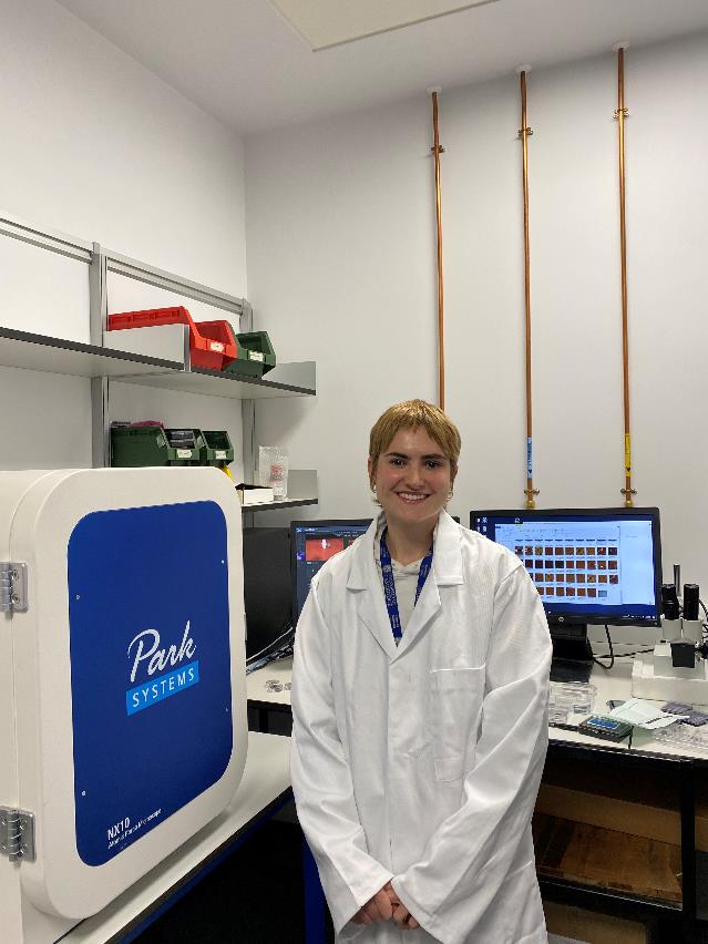
2. Why is the Park AFM important for your research?
The Park AFM has been central in my research, as it enables to study the morphology of protein aggregates with nanometer-scale resolution. Protein assemblies are notoriously difficult to study, due to their small size, typical low abundance in solution, and dynamic nature. AFM is thus very well-suited to capture localized structural properties.
3. What features of Park AFM are the most beneficial and why?
The robust nature of the Park NX10 instrument makes it easy to achieve high-resolution images of few nanometer-sized protein structures, such as individual monomers. As we are often studying small changes between heterogenous samples, we need to take many images to ensure that we are accurately representing our biomolecules. The Park is very strong in this respect, as it can scan reliably for days with minimal user input. The phase-readout also allows us to monitor how ‘hard’ we are scanning our sample, meaning that we can gently image our soft biological samples without damaging them.