


Imaging the potential of molecule aggregates via sideband KPFM
Ilka M. Hermes, Andrea Cerreta
Park Systems Europe GmbH, Mannheim, Germany
The work function is a material characteristic property that can be used to distinguish single constituents in composites or differentiate between sample and substrate. Kelvin probe force microscopy (KPFM) can image the work function distribution of surfaces with a nanometer resolution for a known tip work function. Here, we present the sideband KPFM, newly implemented on Park Systems research atomic force microscopes (AFMs). Sideband KPFM improves the potential sensitivity and spatial resolution significantly and thereby increases the accuracy and reliability of KPFM measurements.
Semifluorinated alkanes consist of two chain segments – one (CF2)xF and one (CH2)yH block. FxHy are known to self-assemble in water and on solid substrates in various morphologies. Therefore, studying of semifluorinated alkanes like F14H20 adds to the general understanding of self-assembly.1 For nanoscale visualization of self-assembled F14H20 structures Kelvin probe force microscopy (KPFM) is ideally suited, since the electric dipole of F14H20 leads to a significant surface potential difference between F14H20 and the substrate.2
KPFM, a scanning probe microscopy technique, captures the surface topography simultaneously to the surface potential. For KPFM, an oscillating conductive cantilever scans the surface, while applying an AC voltage to detect changes in the electrostatic force between tip and sample caused by local variations of the surface potential.3 To minimize the detected electrostatic force, a DC bias counteracts the contact potential difference between tip and sample at each point of the scan. Based on the applied DC bias the surface potential distribution of the sample is reconstructed in the KPFM signal. If the work function of the conductive tip is known, the potential distribution can be converted into the work function distribution of the sample. For the resolution and accuracy of the surface potential in KPFM, the detection method of the electrostatic force is decisive.
In off-resonance KPFM, the AC voltage modulates the electrostatic force at a frequency far from the resonance of the cantilever, used for topography imaging (Figure 1a). The force is then detected via the oscillation amplitude at the AC frequency. By applying a DC bias that matches the potential difference between tip and sample the amplitude at the AC frequency and therefore the electrostatic force is nullified. However, the dependence of the KPFM signal on the long-ranged force lowers the sensitivity of the measurement, because non-local interactions between sample and cantilever can superimpose on the local signal.3
For sideband KPFM, we apply a low-frequency AC voltage (2-5 kHz) to modulate the electrostatic force gradient. The modulated force gradient introduces frequency sidebands left and right of the cantilever resonance (Figure 1b). Analogous to off-resonance KPFM, sideband KPFM nullifies the amplitude of these sidebands by applying a DC bias matching the potential difference. By detecting the short-range force gradient instead of the force, long-range crosstalk decreases and the lateral resolution and local potential sensitivity improve significantly.3
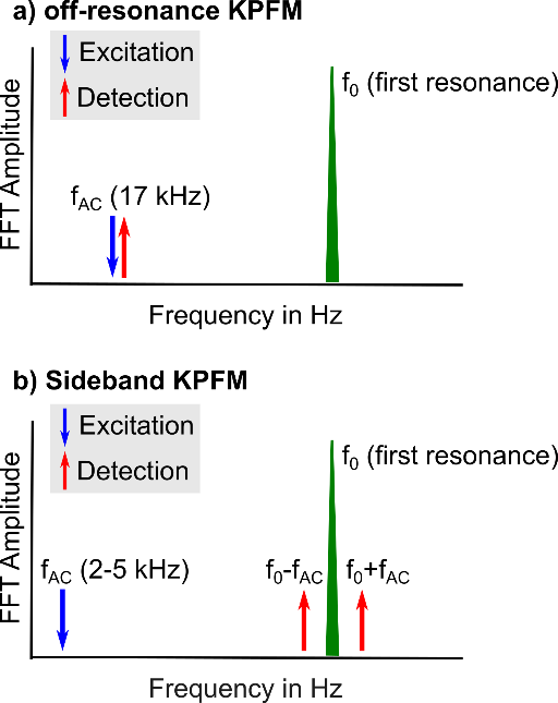
Figure 1: Schematic frequency spectra for off-resonance KPFM in a) and sideband KPFM in b).
Sideband KPFM on electrode array
To test the potential resolution and accuracy of sideband KPFM before we measure on the F14H20, we applied different voltages (0 V and -2 V) to adjacent electrodes on a gold electrode array (Figure 2a). The overlay of the sample topography and the sideband KPFM potential in Figure 2b illustrates the different potential that was detected on both electrodes: while the left electrode shows a bright potential contrast of around 0 V, the right electrode features a dark potential contrast of around -2 V. The more detailed measurement analysis in Figure 2c includes a line profile across the potential image. Here, we found that the measured potential is in agreement with the applied voltages. As such, we detected a 2 V difference between both adjacent electrodes as well as a sharp transition from the electrodes to the substrates. Therefore, we demonstrated that sideband KPFM can capture the full voltage applied to the sample with a high potential sensitivity and spatial resolution.
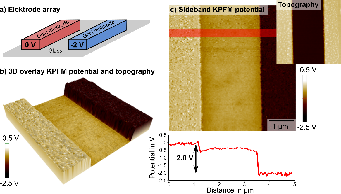
Figure 2: a) Array of gold electrodes with voltages of 0 und -2 V, respectively. b) 3D overlay of sideband KPFM potential and topography illustrates the two different potentials of both electrodes according to the applied voltages. c) Sideband KPFM potential with a line profile across both electrodes along the red line shows that the measured potential agrees with the applied voltages and a high spatial resolution.
KPFM on F14H20 molecule aggregates
For the comparison of sideband KPFM with the more commonly applied off-resonance KPFM, we mapped the surface potential distribution on self-assembled aggregates of semifluorinated Alkanes (F14H20). Here, the electric dipole of the molecules introduce a significant potential offset between the aggregates and the subtrate.2
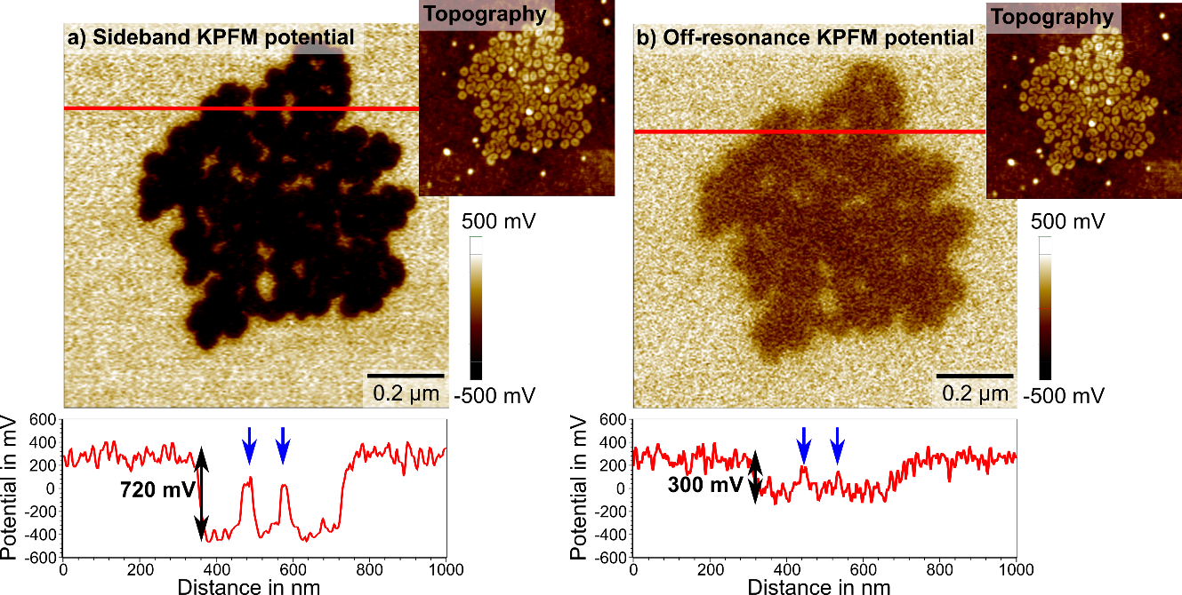
Figure 3: The same F14H20 aggregate was imaged using off-resonance and sideband KPFM. Cross sections (red) show improved lateral and potential resolution of sideband KPFM.
The results of the off-resonance and sideband KPFM measurements showed an improved spatial and potential resolution for sideband KPFM. For sideband KPFM, we observed a potential contrast of 700-750 mV between substrate and F14H20, as well as a defined lateral resolution, imaging even small gaps in the aggregate. Off-resonance KPFM on the other hand showed a potential difference of around 300 mV, indicating a lower local potential sensitivity. Furthermore, the distinct edges captured in sideband KPFM were blurred in off-resonance KPFM, highlighting the superior spatial resolution of sideband KPFM.
The softness of F14H20 molecule aggregates posed an additional challenge to characterization via scanning probe techniques. However, sideband and off-resonance KPFM can be combined with Park Systems’ non-contact mode, thus allowing for stable topography imaging of these soft molecule structures.
Summary
Sideband KPFM, available on all Park’s NX research tools, offers accurate surface potential measurements on soft samples like F14H20 as well as for semiconducting and metallic materials. The dependence on the electrostatic force gradient significantly improves the lateral resolution and potential sensitivity, making sideband KPFM the ideal technique for quantitative surface potential characterization on the nanoscale.
Read the corresponding appnote on Surface Potential Imaging via Sideband Kelvin Probe Force Microscopy
Source
1. Silva, G. M. C., Morgado, P., Lourenço, P., Goldmann, M. & Filipe, E. J. M. Spontaneous self-assembly and structure of perfluoroalkylalkane surfactant hemimicelles by molecular dynamics simulations. Proc. Natl. Acad. Sci. 116, 14868 LP – 14873 (2019).
2. Silva, G. M. C., Morgado, P., Lourenço, P., Goldmann, M. & Filipe, E. J. M. Spontaneous self-assembly and structure of perfluoroalkylalkane surfactant hemimicelles by molecular dynamics simulations. Proc. Natl. Acad. Sci. 116, 14868 LP – 14873 (2019).
3. Zerweck, U., Loppacher, C., Otto, T., Grafström, S. & Eng, L. M. Accuracy and resolution limits of Kelvin probe force microscopy. Phys. Rev. B 71, 125424 (2005).