


The 8th Multifrequency AFM Conference will take place in a virtual format on October 27-30. Park Systems is proud to be a sponsor of the conference!
The Multifrequency AFM conference series aims to provide the environment where the experts and the newcomers in the force microscopy and nanomechanics fields share knowledge, expertise and ideas about instrumentation, methodologies and theoretical aspects on advanced atomic force microscopy.
This year the conference will focus on such topics as high speed AFM, nanomechanics, bimodal AFM, high resolution imaging of soft matter, 3D imaging of solid-liquid interfaces, cantilever design and dynamics and multifrequency methods.
Join our 2 exciting talks:
1. "Simultaneous vertical and lateral resonance tracking PFM on ferroelectric" presented by Principal Scientist - Ilka Hermes on Thursday, 29th October at 12:30 pm,
2. "Capturing the full potential: Surface potential imaging of soft structures via sideband KPFM" presented by Applications Scientist - Andrea Cerreta on Wednesday, 28th October at 16:10 pm.
Please see the abstracts below.
- Event Dates: 27 – 30 October 2020
- Venue: Colegio Oficial de Odontólogos y Estomatólogos de la Primera Región, Madrid
Link: https://wp.icmm.csic.es/multifrequency-afm/
_____________________________________________________________________________________________________________________
Simultaneous vertical and lateral resonance tracking PFM on ferroelectric thin films
Even prior to the rise of perovskite solar cells, functional ferroelectric thin films have been investigated for their potential application in electronic and optoelectronic devices. For instance, the bulk photovoltaic effect based on ferroelectricity promises above band gap photovoltages for photovoltaics. Furthermore, conductive domain walls between ferroelectric domains could act as charge carrier pathways lowering recombination rates and, thus, increase the charge collection in electronic devices.1–3
To correlate ferroelectric effects in domains on electronic and optoelectronic properties in ferroelectric functional materials vertical and lateral domains should be visualized with a high spatial resolution. The local piezoelectric information becomes available via piezoresponse force microscopy (PFM). For thin films, the weak piezoresponse can be enhanced by driving the electrical excitation of PFM close to the vertical (deflection) or lateral (torsion) contact resonance. Since the contact resonance depends on a consistent tip-sample contact a high surface roughness often introduces topographic crosstalk.4 Dual frequency resonance tracking (DFRT) improves the stability of the resonance enhancement.5 Here, we demonstrate our capabilities to capture the out-of-plane and in-plane DFRT piezoresponse simultaneously, by driving the electrical excitation of the cantilever at the contact resonance of the vertical deflection, as well as the torsional resonance on ferroelectric thin films, including bismuth ferrite and lead zirconium titanate.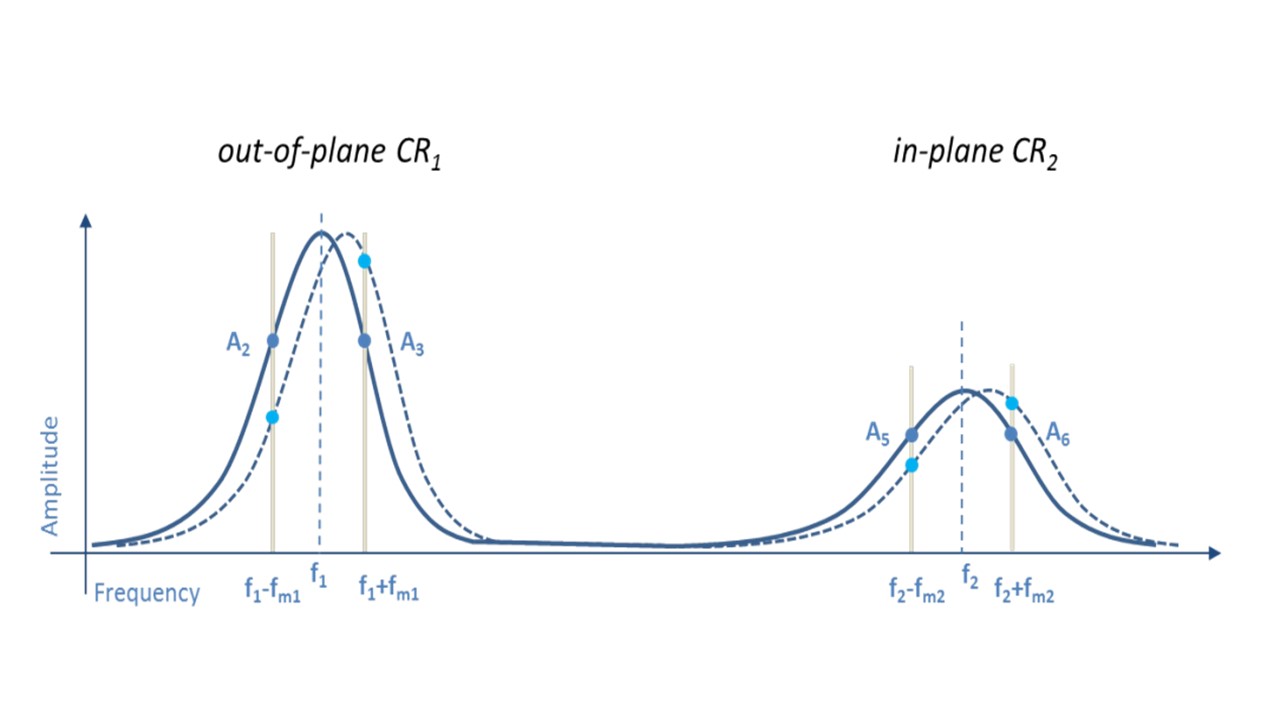
Figure 1: Frequency spectrum of cantilever in contact including the out-of-plane vertical resonance CR1 and the in-plane lateral resonance CR2. The sidebands used for the resonance tracking (A2 and A3 for the vertical resonance and A5 and A6 for the lateral resonance) are visualized in grey.
____________________________________________________________________________________________________________________
Capturing the full potential
Surface potential imaging of soft structures via sideband KPFM
Andrea Cerreta1, Ilka Hermes1
1Park Systems Europe GmbH, Mannheim, Germany
Semifluorinated alkanes consist of two chain segments – one (CF2)xF and one (CH2)yH block. FxHy are known to self-assemble in water and on solid substrates in various morphologies. Therefore, studying of semifluorinated alkanes like F14H20 adds to the general understanding of self-assembly.1
For nanoscale characterization of soft, self-assembled F14H20 structures on solid substrates Kelvin probe force microscopy (KPFM) is ideally suited, since the electric dipole of F14H20 leads to a significant surface potential difference between F14H20 and the substrate.2 In KPFM, a conductive cantilever scans the sample, while applying an AC and a DC voltage to detect changes in the electrostatic force between tip and sample caused by local deviations of the surface potential.3
Here, we compared KPFM measurements of self-assembled F14H20 structures imaged by two techniques: Off-resonance and Park’s newly implemented sideband KPFM. We found a significant improvement in the potential resolution for sideband KPFM.
KPFM on F14H20
For the resolution and accuracy of the surface potential in KPFM, the detection method of the electrostatic signal is decisive. In off-resonance KPFM, the AC voltage modulates the electrostatic force at a frequency far from the resonance of the cantilever (~17 kHz). The force is detected via the oscillation amplitude at the AC frequency, which is nullified by applying a DC bias matching the potential difference between tip and sample. However, the detection of the long-ranged force can lower the sensitivity.3
For sideband KPFM, we apply a low-frequency AC voltage (2-5 kHz) to modulate the electrostatic force. The modulated electrostatic force gradient introduces frequency sidebands left and right of the cantilever resonance. Sideband KPFM nullifies these sidebands by applying a DC bias, which matches the potential difference. By detecting the force gradient instead of the force, long-range crosstalk decreases and the lateral resolution and potential sensitivity improve significantly.3
Analyzing the sideband KPFM measurement on a F14H20 aggregate on silicon substrate, we observed a potential contrast of 700-750 mV between substrate and F14H20 as well as a defined lateral resolution, imaging even small gaps in the aggregate. Off-resonance KPFM on the other hand showed a potential difference below 300 mV and a lower lateral resolution.
Summary
Sideband KPFM, available on all Park’s NX research tools, offers accurate surface potential measurements on soft samples like F14H20 as well as for semiconducting and metallic materials. The dependence on the electrostatic force gradient significantly improves the lateral resolution and potential sensitivity, making sideband KPFM the ideal technique for quantitative surface potential characterization on the nanoscale.
References
1. G. M. C. Silva, et al, Proc. Natl. Acad. Sci., 2019, 116, 14868.
2. A. El Abed, et al, Phys. Rev. E, 2002, 65, 51603.
3. U. Zerweck, et al, Phys. Rev. B, 2005, 71, 125424.
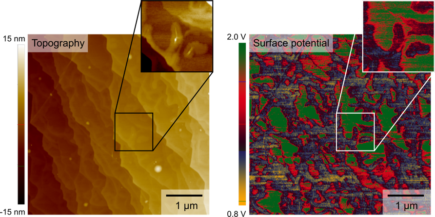
Figure 1:
The same F14H20 aggregate was imaged using off-resonance and sideband KPFM. Cross sections (red) show improved lateral and potential resolution of sideband KPFM.