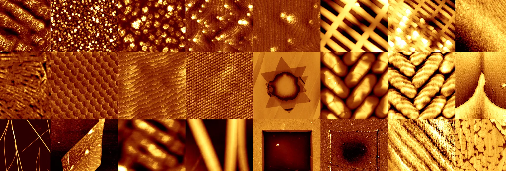
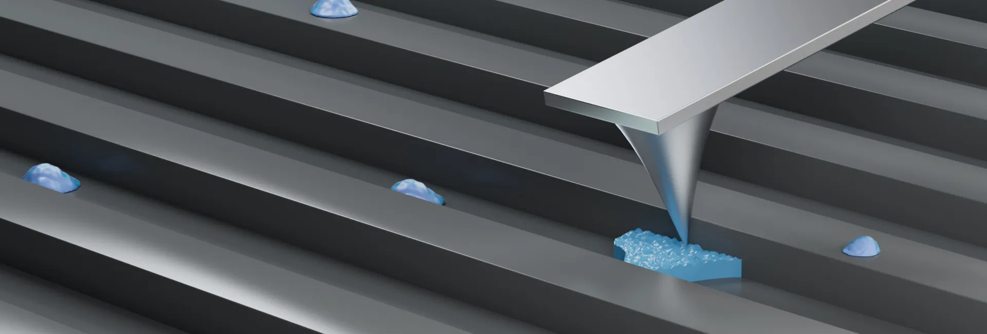
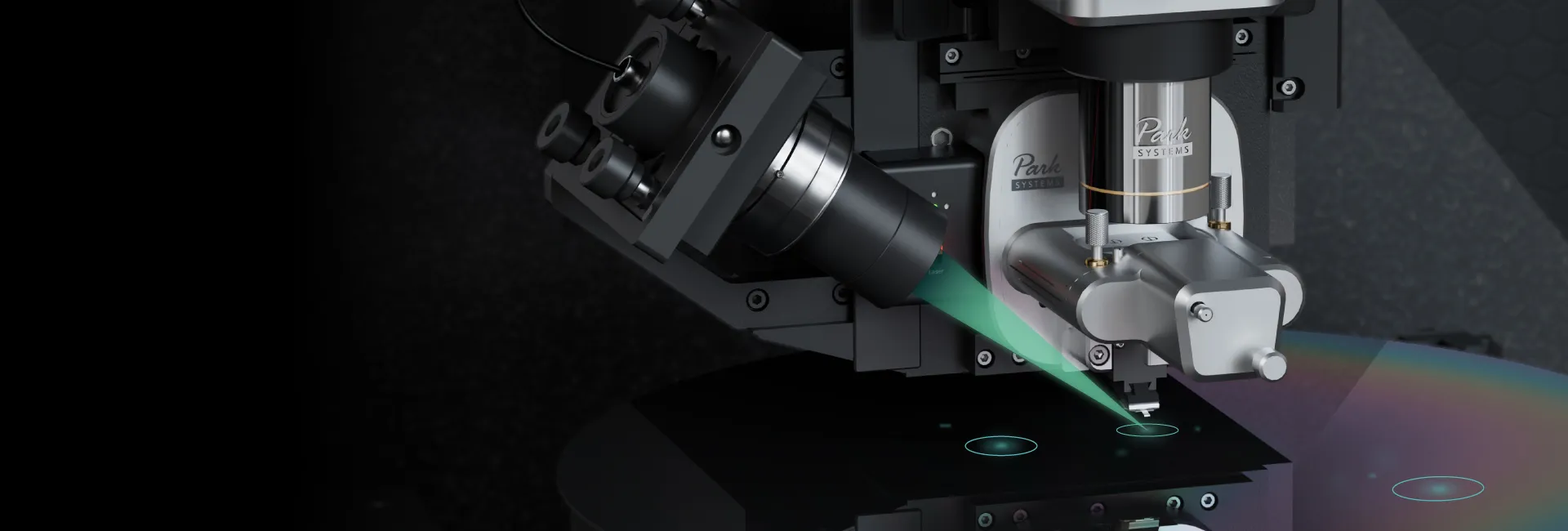
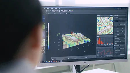
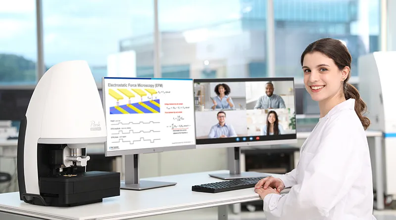
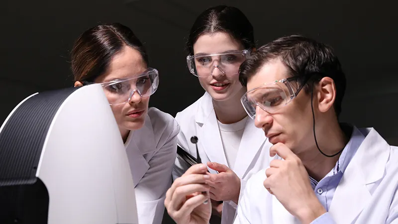
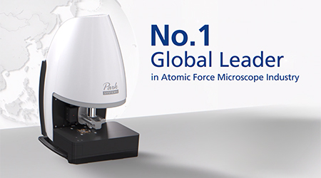
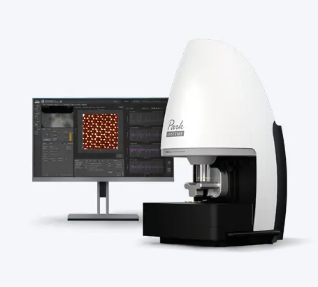
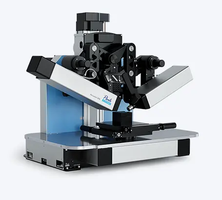
Atomic Force Microscope (AFM) is an indispensable tool in the field on nanotechnology to map nanometre sized features and physical properties of the sample surface. AFM can be used to image different kinds of samples such as polymers, thin films, nano particles, metals, semi-conductors, ceramics, glass, and biological materials etc. Majority of these samples can be imaged under ambient conditions and follow a relatively simpler sample preparation procedure. The sample must be firmly attached to the solid substrate since the AFM tip physically interacts and tracks the sample surface. So the knowledge of sample preparation becomes important to achieve best and accurate AFM images. It is also important to investigate the substate preparation on which the sample can be dispersed and adhered firmly. Thus, AFM imaging fundamentally requires the substrate to be clean and optically flat as possible, the sample affixed on the substrate and substrate also fixed or firmly attached. In this webinar we highlight the procedures of sample preparation for an AFM. These procedures are generally optimised for specific type of samples and if followed correctly can generate high resolution images on an AFM.
- 제품소개
- 연구ᆞ표면분석용 원자현미경
- Small Sample AFM
- Large Sample AFM
- Vacuum Environment AFM
- AFM Probes and Options
- AFM Modes and Techniques
- 인라인 계측용 원자현미경
- AFM for Wafer Fabs
- AFM for Flat Panel Display
- Photomask Repair
- Optical Profilometry
- Nano Infrared Spectroscopy
- Ellipsometry for Thin Film Characterization
- Imaging Spectroscopic Ellipsometry
- Referenced Spectroscopic Ellipsometry
- Brewster Angle Microscopy
- Ellipsometry Accessories
- Surface Inspection Metrology
- 응용기술
- 고객지원
- 이벤트
- 회사소개
- 러닝센터
- NANOacademy
- Lectures
- How AFM Works
- 전문가 코너
- Analyze Cells
- Programs
- Park AFM Scholarship
- 제품소개
- 연구ᆞ표면분석용 원자현미경
- 인라인 계측용 원자현미경
- Ellipsometry for Thin Film Characterization
- Active Vibration Isolation
- Software
- 응용기술
- 고객지원
- 이벤트
- 회사소개
- 러닝센터
- NANOacademy
- Programs
- Resources
Copyright © 2024 Park Systems. All Rights Reserved.


