


Nanomechanical Mode
PinPoint™ Nanomechanical Mode
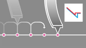
PinPoint™ Nanomechanical mode obtains the best of resolution and accuracy for nanomechanical characterization. Stiffness, elastic modulus, adhesion forces are acquired simultaneously in real-time. While the XY scanner stops, the high speed force-distance curves are taken with well-defined control of contact force and contact time between the tip and the sample. Due to controllable data acquisition time, PinPoint™ Nanomechanical mode allows optimized nanomechanical measurement with high signal-to-noise ratio over various sample surfaces.
Read MoreRead more about accurate Nanomechanical Imaging via PinPoint here
Video: Characterizing Multicomponent Polymer with PinPoint™ AFM
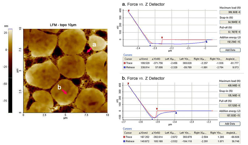
Force-Distance Spectroscopy
Force Measurement of Tip-Sample Interaction

Here, Force distance (FD) spectroscopy is a straightforward and reliable technique to quantitatively study nanomechanical properties such as Young’s modulus and adhesion force on a variety of samples. Therefore, FD spectroscopy has become a fundamental characterization tool in several fields of research, including polymer science, biochemistry, and biology.
Read More
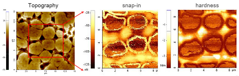
Force Volume Imaging
Force Measurement of Tip-Sample Interaction
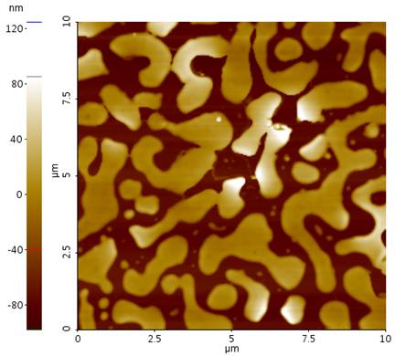
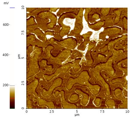
Force volume imaging provides a detailed map of the sample’s material properties by plotting parameters such as stiffness, cantilever snap-in, and adhesion. Parameters extracted from Force Distance (F-D) spectroscopy curves are put into a matrix that quickly and easily allows researchers to gain insights about samples properties.
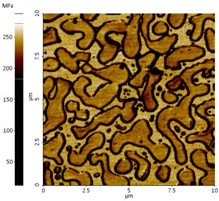
Force Modulation Microscopy (FMM)
Force Amplitude and Phase Imaging of Sample Elasticity
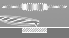
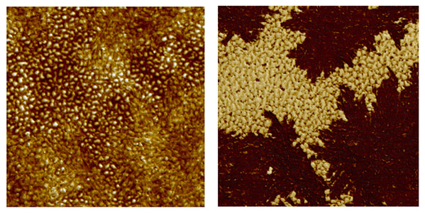
In addition to topographic imaging, Atomic force microscopy (AFM) is routinely used to resolve mechanical properties of various samples for material and life science on the nanometer scale. An established technique to probe nanomechanical properties is Force modulation microscopy (FMM). FMM is based on contact mode AFM with an additional mechanical modulation that is applied to the cantilever during the contact scan.
Read More
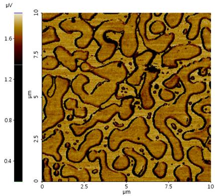
Lateral Force Microscopy (LFM)
Mapping of the Frictional Force
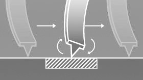
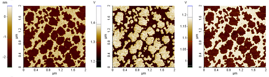
In AFM, frictional properties can be investigated via Lateral force microscopy (LFM). LFM can be used to study differences in material compositions on coating layers, lubricant properties, strength of adhesion on patterned structures and so on.
Read More
Nanolithography
Advanced Vector Nanolithography Using Closed Loop Scan System
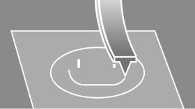
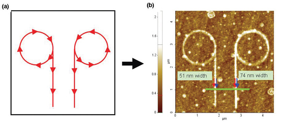
The Nanolithography mode allows you to manipulate and create patterning on the sample surface through applied force or voltage. Tip position for lithography can be easily controlled by importing your own vector drawings or raster (bitmap) images.
Read More
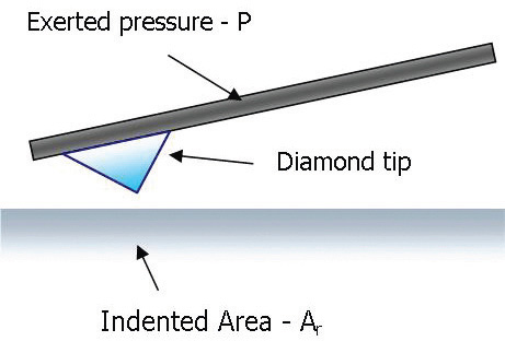
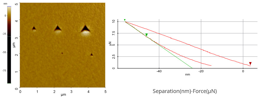
Nanoindentation
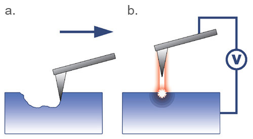
Atomic force microscopy (AFM)-based Nanoindentation quantitatively characterizes local mechanical properties of target specimen. In this technique, a hard AFM indentation tip with known mechanical properties presses against a sample surface until the tip deforms the surface.
Read More
Spring Constant Calibration by Thermal Method
Maintaining the proper spring calibration is critical for AFM force data accuracy. That’s why Park provides spring calibration using the thermal method, giving you accurate readings every time.