


Brian Choi, Bio-application scientist
For more information, please contact app@parksystems.com
Data Reference: Gordon Jung, Application Scientist
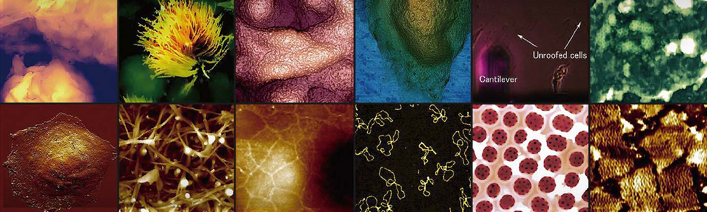
Cell imaging in live status provides critical insight into the fundamental nature of cell and tissue function. Especially importantly, imaging a cell membrane in high resolution and monitoring its dynamics can reveal a variety of cellular processes and cell signaling behavior. To capture the real nature of cell membrane dynamics, it is important that the cell’s physiology is not interrupted by imaging technique in use. Scanning ion conductance microscopy (SICM) is a non-contact surface morphology imaging technique, operating in an ion-containing liquid environment. SICM provides automatic time-lapsed images and shows real-time changes in a cell membrane’s small features during imaging.
- Monitor the dynamics of cell membrane and nanostructures of live cell
- Automatic running mode (ARS) visualizes and records morphology changes of live cell membrane
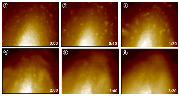 Cell Membrane of C2C12
In this study, the cell membrane morphology of living C2C12 (myoblast) and HeLa cells, as well as the dynamics of those membranes, were captured by SICM over time. The continuous imaging of cell membranes unveils the detail morphological physiology of C2C12 and HeLa cells by showing outward cellular extensions, such as microviili, covering the plasma membrane. It is clearly observed that the microvilli pops down crossing the cell membrane (C2C12), and those cellular extensions are transforming its structure and moving the position dynamically in the lateral direction as time passes (HeLa cell).
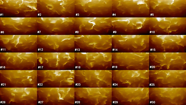 Cell Membrane of Live Hela
Park Cell Analysis Systems
|
|
|
|
| Park NX12-Bio | Park NX10 | Park XE7 | |
| Scanning Ion Conductance Microscopy (SICM) | |||
| Atomic Force Microscopy (AFM) with liquid probe hand | |||
| Inverted Optical Microscopy (IOM) | |||
| Live Cell Chamber |