


Brian Choi, Bio-application scientist
For more information, please contact app@parksystems.com
Reference: Noemi Rozlosnik, et al. (2006), Nanomechanics of Single Muscle Fibers by AFM,
International Nano-Conference(ICN+T), Basel (CH)
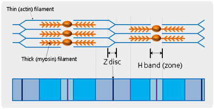
Muscle fiber is composed of skeletal muscle, organized with multi-nucleated muscle cells or myofibers. All individual muscle fibers are arranged into bundles and then finally forms the muscle belly. The basic unit of muscle fiber is called, a ‘sarcomere’ and is defined being between neighbouring Z-discs in a muscle fiber. In the middle of a sarcomere, there is a paler region called the ‘H-band (zone)’ in which muscle relaxation and contraction occur. Utilizing atomic force microscopy (AFM) and its force distance spectroscopy, it is possible to study the mechanical property of the detail units of sarcomere. After identifying a specific feature of biological objects from the high resolution AFM topography, cantilever tip can be brought to the feature and indent it in accurate force control at nanonewton scale.
- Study the mechanical property of the detailed units of sarcomere using AFM and its force distance spectroscopy.
- AFM topography and XY scanner enables the indentation of the cantilever tip at the desired position on the sarcomere with nanometer precision.
- The stiffness of Z disc and H band are accurately calculated and distinguishable in nanonewton range.
Structure of Muscle Fiber (Sarcomere)
The mechanical property of a sarcomere can be analyzed quantitatively by performing the force distance spectroscopy data wherein the force distance curve is acquired on the muscle fiber. Depending on each indenting positions, the corresponding mechanical property can be varied so that the cantilever deflections are recorded differently. The deflection differences are distinguishable, at nanonewton scale, by calculating the ‘stiffness (the angle of loading curve)’ in the recorded force distance curves.
Muscle Fiber Stiffness Measurement by Force-Distance Spectroscopy of AFM
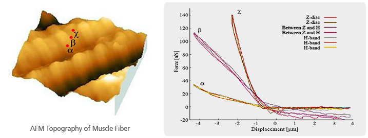
The muscle fiber topography imaged with Park AFM showed the typical morphology of a sarcomere as well as the feature of Z disc and H band. Controlling XY scanner position to one of the sarcomere units, three force distance curves were acquired - H band (α), between H band and Z disc (β), and Z disc (χ). As calculated, the stiffness of H band (α) is about 10 nN/μm, about 45 nN/μm for ‘between H band and Z disc (β)’ and about 110 nN/μm for Z disc (χ). It is obvious that the Z disc (χ) 11 times higher in stiffness than the H band (α).
Park Cell Analysis Systems
|
|
|
|
| Park NX12-Bio | Park NX10 | Park XE7 | |
| Scanning Ion Conductance Microscopy (SICM) | |||
| Atomic Force Microscopy (AFM) with liquid probe hand | |||
| Inverted Optical Microscopy (IOM) | |||
| Live Cell Chamber |