|
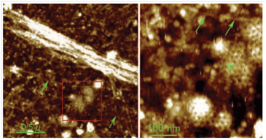
Researchers at Nagoya University, headed by Professor Jiro Usukura, can unroof a cell membrane by ultra-sonication and expose the inner side or cytoplastmic surface of the cell membrane. Park XE-Bio with True Non-Contact ModeTM successfully imaged the very soft and high resolution details of intracellular cytoskeleton and the cytoplasmic surface of the cell membrane under aqueous condition.
Many actin filaments were found to form a complex meshwork on the cytoplasmic surface of the membrane. Characteristic periodic striations of about 5 nm formed by the assembly of G-actin were detected along actin filaments at higher magnification. Microtubules were also easily detected and were often tethered to the membrane surface by fine filaments. Furthermore, clathrin coats on the membrane were clearly visualized for the first time in water by atomic force microscope (AFM).
Automatic Defect Review (ADR) for hard disk media and substrates provides a 500-800% gain in throughput compared to current manual methods. With automated reference marker detection, stage mapping, transfer of Candela defect maps, survey & zoom-in imaging, and profiling of imaged defect types, ADR allows automated defect identification and analysis with a success rate of over 95%. Also, with Park Systems’ True Non-contact mode it increases tip life significantly which enables, with a single tip, the complete full cycle run of samples that contains 100s of defects.
Click here to read the Jouranl of Electron Microscopy pager.
|
News and Events
 |
NX20 : World's Most Accurate AFM for Failure Analysis and Quality Assurance.
Santa Clara, California, November 19, 2012 - Park Systems introduces the NX20, a high-end, large sample atomic force microscope (AFM) for failure analysis (FA) and quality assurance (QA) laboratories that will benefit from the best available accuracy and reliability. The NX20 is the newest model of the NX product line with the world’s most accurate AFM. Designed for the needs of FA and QA in the hard disk drive and semiconductor industries, the NX20 stands out for its unmatched non-contact mode that guarantees long-running probe tip sharpness. That’s the key to the highest accuracy and repeatability for roughness measurement and defect review. It’s also key to high productivity and the lowest AFM lifecycle costs.
“Our technology has been proven in the most demanding applications for nanoscale measurements in industry,” said Dr. Sang-il Park, the founder and CEO of Park Systems. “NX20 builds on Park's long history of technology leadership in AFM accuracy and its reputation as the nanotechnology solutions partner to research and industry. Our True Non-Contact Mode preserves the tip for much longer tip life and provides the most accurate measurements and images available for the most critical analysis requirements.”
Click here for the full NX20 article.
|
|
Emphor Fzco appointed distributor for Park Systems in the United Arab Emirates.
Santa Clara, California, October 1, 2012 - Park Systems, the atomic force microscopy leader, and Emphor free zone company (Dubai, UAE), a leading technology integrator and distributor in the Middle East, announced today a new distribution agreement enabling Park Systems to extend its global presence and promote its atomic force microscopy (AFM) products in the UAE, Qatar, Bahrain and Oman.
Emphor operates under the umbrella of Centena, a free zone holding company with headquarters in Dubai, UAE. Since its founding in 1980, the Centena group has a rich commercial history of partnering with leaders in their respective businesses from across the globe. Under its operational holdings are Tensosys (maritime electronics), ScreenCheck (smart card solutions), Maritronics (maritime systems integration) and Emphor.
Clcik here for the full news article.
Park True Non-Contact ModeTM Uncovers the Secrets of Inside Cell Membrane.
Santa Clara, California, December 10, 2012 – Park Systems XE-Bio, the leading atomic force microscopy (AFM) system for cell biology imaging, was featured in the Journal of Electron Microscopy [October, 2012] in an article entitled “Use of the unroofing technique for atomic force microscopic imaging of the intra-cellular cytoskeleton under aqueous conditions.” The article was the Editor’s Choice for the bimonthly journal and an image from the article taken by the Park XE-Bio (the cytoplasmic surface of the ventral cell membrane) was the journal’s cover image.
The article describes the research of Professor Jiro Usukura and his team at Nagoya University EcoTopia Science Institute (Japan). Using ultrasonication, the researchers unroofed a cell membrane (removed the top layer) to expose the inner side or cytoplasmic membrane. Once exposed, the Park XE-Bio atomic force microscope was able to image the very soft and high-resolution details of the underlying intracellular cytoskeleton and the cytoplasmic surface of the cell membrane. The XE-Bio can do this very delicate imaging, even in an aqueous environment, thanks to its True Non-Contact ModeTM where the tip of the AFM probe can collect extremely accurate imagery data without making contact with the surface of the sample. This mode preserves the physical integrity of the sample while extending the life of the AFM probe tip.
Click here for the full news article.
|