Brian Choi, Bio-application scientist
For more information, please contact app@parksystems.com
Reference: Jiro Usukura, et al. (2012), Use of the unroofing technique
for atomic force microscopic imaging of the intra-cellular cytoskeleton under aqueous conditions,
Journal of Electron Microscopy 61(5): 321–326
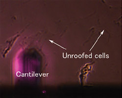
Atomic force microscopy (AFM) is a useful method for acquiring sub-cellular molecule image of biological objects. There are advantages of AFM to biology study. It has high resolution (<10 nm) that enables the resolving of small molecules like DNA and protein. Another advantage is its ability to acquire high resolution images in liquid environment so the physiological living activity of biological features can be visualized under natural conditions. Unroofing technique is a cell treatment method from which the upper membrane of live cell are removed clearly so that intra-cellular cytoplasmic surface of membrane is exposed in aqueous conditions.
- AFM’s high resolution (<10 nm) imaging, capable in aqueous conditions provides the chance to observe sub-cellular molecule in vivo.
- Acquire the high resolution image of intracellular cytoskeleton and clathrin-coated pits in buffer with AFM, employing unroofing technique.
Unroofing Technique for Sub-cellular Molecule Imaging with AFM

Employing the unroofing technique, atomic force microscopy (AFM) enabled the high resolution imaging of the intracellular cytoskeleton and the cytoplasmic surface of the cell membrane under aqueous conditions.
Intra-cellular Cytoskeleton and Clathrin-coated Pits Image of AFM
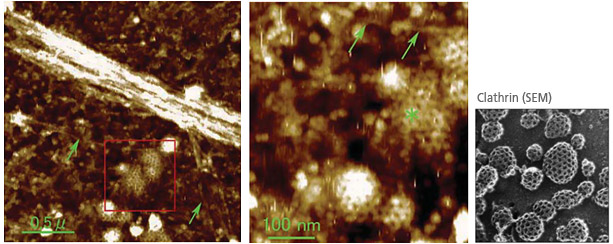
Many actin filaments were found to form a complex network on the cytoplasmic surface of the membrane. Furthermore, clathrin coats on the membrane were clearly visualized for the first time in liquid by AFM. Although the resolution of these images is lower than the one from scanning electron microscopy, the measurement capabilities of the AFM is more biologically relevant. AFM imaging in aqueous conditions is an important capability for investigating various sub-cellular molecules such as intracellular cytoskeleton and clathrin-coated pits in the native, aqueous state. It holds the key of many undiscovered biological secrets.
Park Cell Analysis Systems
|
|
|
|
| Park NX12-Bio | Park NX10 | Park XE7 | |
| Scanning Ion Conductance Microscopy (SICM) | |||
| Atomic Force Microscopy (AFM) with liquid probe hand | |||
| Inverted Optical Microscopy (IOM) | |||
| Live Cell Chamber |