


Bio Atomic Force Microscope: Liposomes and Vesicles
AFM imaging as a Tool to Visualize Liposomes and Vesicles
Lipid bilayer formed by the fusion of phospholipid vesicles is invaluable to researchers in the study of the characteristics and behavior of membrane-bound proteins, cell membrane and signal transduction. Phospholipid vesicles or liposomes, however, are also important to the study of secretory vesicles.
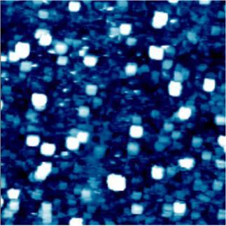
Figure 1. AFM image of liposomes in liquid imaged with the XE-100 (2 μm size scan) Non-Contact mode.
A vesicle is a relatively small and enclosed compartment, separated from the cytosol by at least one lipid bilayer. The vesicle is the simplest organelle in the cell. These vesicles are used for chemical reaction chambers, enzyme storage, and transporters.
Vesicle trafficking is a common source of manifestation of many human pathologies. Dysfunction in vesicle trafficking in targeting and delivery of specific proteins to the cell surface have serious consequences and have been implicated in diseases such as Alzheimer’s, cystic fibrosis, type II diabetes and polycystic kidney diseases, just to name a few.
AFM can be utilized to study the size dynamics of vesicles in isolated preparations in liquid. The swelling dynamics of vesicles and interaction with target membranes can be visualized with high resolution.
Phospholipid vesicles or liposomes can mimic secretory vesicles such as zymogen granules or synaptic vesicles by incorporating vesicular proteins. Fundamental studies on vesicular membrane ligand-receptor interaction, membrane adhesion and fusion, and numerous other biophysical phenomena can be studied.
By using specific substrates such as titanium oxide, which resists lipid absorption, AFM can image liposomes. Non-Contact mode of Park AFM is specially useful for imaging ultra-soft materials like liposomes in liquid.
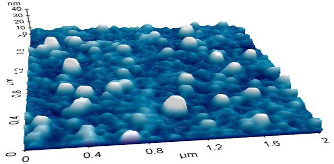
Figure 2. 3D rendering of AFM images of lliposomes in liquid with the XE-100 (2 μm size scan) Non-Contact mode.
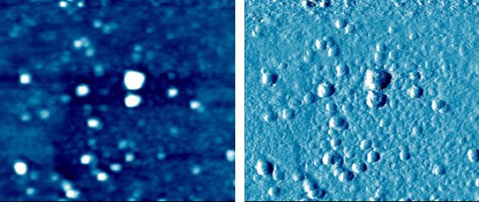
Figure 3. AFM topography and error images of lliposomes in liquid with the XE-100 (2 μm size scan) Non-Contact mode.