


Useful to Study Morphology and Physical Properties of Cells Derived from Human Brain Tumor
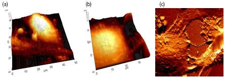
Figure 1. AFM Images of human astrocytoma cells. (a) Topography image (50 μm scan size), (b) topography image (40 μm scan size) and (c) error image (55 μm scan size).
Human astrocytes, one type of glial cells, are major components of the brain tissue, which supports neurons and blood vessels in the brain. Astrocytoma cells are derived from brain tumor whose origin is in the astroglial lineage.

Figure 2. AFM images of human astrocytoma cells. (a) Topography image (70 μm scan size), (b) FMM amplitude image (70 μm scan size), and (c) living cell in PBS (33 μm scan size).
Figure 1 and 2 show AFM images of human astrocytoma cells, U373 MG in PBS. The cells were cultured in DMEM supplemented with 10% FBS in 35 φ T/C dish. Before imaging, the cells were washed with PBS twice and fixed with 1% glutaraldehyde and 1% paraformaldehyde in PBS. Then, the cells were washed with PBS once more and the dish was filled with 3mL PBS.
The images were obtained using contact mode AFM in liquid. A gold-coated silicon nitride cantilever was used to image the sample. Its spring constant is approximately 0.06 N/m.
Remarkably, the center image in Figure 2 shows the amplitude image of FMM (Force Modulation Mode). Fine details of cell surface were displayed in the figure. The image in Figure 2 (c) is taken from living astrocytoma cell in PBS.
It is also possible to investigate physical properties of cells using FMM or a F-d curve. Functionalizing the AFM tip with antibodies of the specific receptor existing on the cell surface will allow the user to obtain antibody recognition images of the cells.
The Park AFM is expected to become a useful tool for studying morphology and physical properties of cells.