


Park SPM Modes
Get the data you need with Park’s selection of scanning modes
Park AFM features a comprehensive range of scanning modes that can collect a wide array of data types accurately and efficiently. From the world's only True Non-Contact™ mode that preserves tip sharpness and sample integrity to advanced Magnetic Force Microscopy, Park Systems offers the most innovative, accurate modes in the AFM industry.
Standard Imaging Mode
Park Systems offers some of the most innovative standard imaging modes in the AFM industry. Park AFM's True Non-Contact™ mode is the world's only truly non-contact AFM scanning mode while our standard scanning mode is among the most accurate available on the market.
True Non-Contact Mode
Park AFM features the only true non-contact standard imaging mode. This mode preserves tip sharpness and sample integrity for better and more accurate scans with less minimal maintenance required.
When True Non-Contact™ mode is activated, a piezoelectric modulator vibrates a cantilever at a small amplitude and fixed frequency near the intrinsic resonance of the cantilever. As the tip approaches the sample, the van der Waals attractive force between the tip and sample influences the amplitude and phase of the cantilever's vibration. These amplitude and phase changes are monitored by the patented Z-servo feedback system of the Park AFM series, which also maintains a few nanometers tip-surface distance without damaging the sample surface or tip.
Specifications :
Tip-sample distance : 3 nm (typical)Cantilever oscillation frequency : 1 - 600 kHz
Cantilever oscillation amplitude : 1 - 2 nm (typical)
Phase Imaging
Phase Imaging with Park AFM provides an accurate measurement of mechanical properties, especially for highly sensitive samples whose topography is best measured using True Non-Contact™ mode. Phase data that contains the mechanical property of the sample is acquired by monitoring the phase lag between the signal that drives the cantilever oscillation and the cantilever oscillation output signal.
Specifications :
Phase resolution :0.005°Lateral Force Microscopy (LFM)
Lateral Force Microscopy with Park AFM provides information on both surface friction and topography by measuring the cantilever deflection in both horizontal and vertical directions. The data acquired in LFM with Park AFM is very useful for studying a sample whose surface consists of inhomogeneous compounds.
Contact Mode
The standard mode of Park AFM is Contact Mode in which the cantilever comes in contact with the surface and the cantilever deflection is measured to produce highly accurate topographic data.
Specifications :
Tip-sample distance control :ContactChemical Properties
Chemical Force Microscopy (CFM)
Park AFM’s Chemical Force Microscopy accurately measures the sample’s chemical properties by using functionalized tips. This technique can also be used in studying simple or complex chemical structures such as proteins and DNA.
Electrochemical Microscopy (EC-AFM)
In Park AFM’S EC-AFM the nanoscale structures of electrochemical reactions on the electrode surface can be observed. Users typically perform voltammetry and corrosion experiments using an electrochemistry cell and a choice of potentiostat/galvanostat depending on the electrochemical application of interest.
Dielectric-Piezoelectric
Park Systems offers a selection of advanced modes that can provide not only topographic imaging but also characterization measurements with respect to the dielectric and piezoelectric property of the sample. These modes include Enhanced Electric Force Microscopy (Enhanced EFM), Dynamic Contact Electric Force Microscopy (DC-EFM), and Piezoelectric Force Microscopy (PFM).
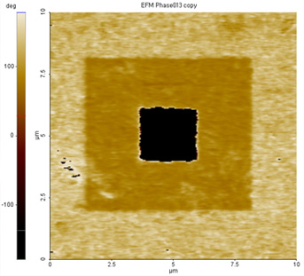
Enhanced Electric Force Microscopy (EFM)
The Enhanced EFM mode of Park AFM offers four separate options: Standard EFM, Dynamic-Contact EFM (DC-EFM), Piezoelectric Force Microscopy (PFM), and Kelvin Probe Force Microscopy (KPFM). The movement of a cantilever is influenced by the electric force between cantilever and sample; cantilever displacement can be analyzed for potential, charge, or electric domains.
Specifications :
Bias range : - 10 V - +10 VDynamic Contact EFM (DC-EFM)*
DC-EFM is capable of extremely high definition EFM results. Patented by Park Systems, DC-EFM actively applies an AC voltage bias to the cantilever and detects the amplitude and the phase change of the cantilever modulation with respect to the applied bias. DC-EFM provides the ability to monitor the second harmonic of the modulation which can be compared to the capacitance of a sample and enhances the electric force signal from the background intermolecular force.
Specifications :
Bias range : - 10 V - +10 VAC bias frequency : 0 - 100 kHz (1 Hz frequency resolution)
US Patent No : 6185991
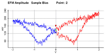
Piezoelectric Force Microscopy (PFM)*
Park Systems’ patented PFM mode accurately measures electric domain structures such as polarity in ferroelectric or piezoelectric samples. This mode includes independent control of an applied AC and DC bias, and local amplitude/phase vs. DC bias spectroscopy.
Specifications :
AC bias frequency : 0 - 100 kHzPhase resolution : 0.005°
DC Bias : - 10 - +10 V (-2kv - +2kV, optional)
US Patent No : 6185991
Piezoelectric Response Spectroscopy
The Piezoelectric Response Spectroscopy mode of Park AFM measures the local amplitude/phase response to a DC bias between tip and sample surface. The polarity of local piezoelectric domain switches depend on the sign and amount of applied voltage.
Specifications :
External high voltage kit requiredDC Bias : -2kV ~ +2kV
Electrical Properties
Park Systems offers a range of electrical scanning modes that can be used by engineers and researchers for highly accurate data measurement on conductance, sample resistance, and other electrical and topographic properties.
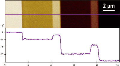
Conductive AFM
Conductive AFM mode with Park AFM provides accurate electrical conductivity information for material processing and characterization of electrical defects in nanomaterials such as nanotubes, conductive polymers, and semiconductors. It can also be used for detailed investigations of a sample’s properties by measuring leakage current and I/V curves of the sample.
Scanning Capacitance Microscopy (SCM)
SCM mode with Park AFM provides doping concentration information over the sample surface by measuring the capacitance change between tip and sample. The module enables a variable resonator frequency, which allows for a wide RF bandwidth capable of monitoring a large range of doping concentrations by selecting the most sensitive frequency of the resonator for a specific doping range.
Specifications :
Frequency range : 600 MHz - 1500 MHz, selectable by software in ~0.1 MHz resolutionFrequency resolution : 2 kHz
Capacitance sensitivity : < 1 aF
SCM Phase resolution : : 0.005°
Scanning Spreading Resistance Microscopy (SSRM)
SSRM mode with Park AFM precisely measures the local resistance over a sample surface by using a conductive AFM tip to scan a small region while applying a DC bias.
Specifications :
Bias range : -10 V - +10 VCarrier concentration : 1015 - 1020 /cm3
Lateral resolution : < 10 nm
Scanning Tunneling Spectroscopy (STS)
STS mode with Park AFM provides current-voltage (I/V) spectroscopy data at user-defined points which can then be used to analyze the local electronic states of the sample.
Specifications :
Batch measurement on user defined points (max.128 x 128 points)Applicable bias range: -10 V to 10 V
Current resolution: < 0.1 pA (ULCA option)
Time-resolved Photocurrent Mapping (Tr-PCM)
Tr-PCM mode with Park AFM measures the photoelectric response to time-resolved illumination without interference from unwanted light sources, including the feedback laser. This mode features a laser illumination module and acquisition and analysis software.
Specifications :
Electric current resolution: 0.03 nAAcquisition time resolution: 20 µsec
Automatic analysis of life-time from photocurrent curves
Force Measurement
Park Systems offers a range of force measurement modes that can be used to easily analyze the sample’s material properties and ensure accurate measurement each time with its unique spring calibration.
Force-Distance Spectroscopy
Force-Distance Spectroscopy mode with Park AFM provides mechanical property information by measuring the vertical force that the tip applies to the surface and the sample displacement while the probe is brought into contact with the surface of the sample at a specific location. It can also be used to analyze surface contaminants’ viscosity, lubrication thickness, and local variations in the elastic properties of the surface.
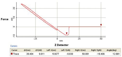
Specifications :
Batch measurement on user specified pointsForce volume image: maximum 128 × 128
Force resolution: < 10 pN
Force Volume Imaging
Force volume imaging with Park AFM provides a detailed map of the sample’s material properties by plotting parameters such as stiffness, cantilever snap-in, and adhesion. Parameters extracted from Force Distance (F-D) Spectroscopy curves are summarized into a matrix that quickly and easily allows researchers to gain insights about samples properties.
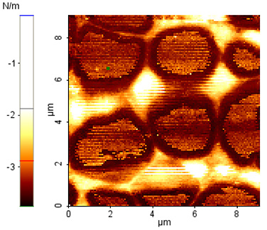
Specifications :
Up to 256 × 256 pointsAutomatic calculation of various F-D parameters (stiffness, snap-in, adhesion)
Analysis software includes batch analysis and export
Spring Constant Calibration by Thermal Method
Maintaining the proper spring calibration is critical for AFM force data accuracy. Hence, Park AFM provides spring calibration using the thermal method, giving you accurate readings every time.
Specifications :
Automatic detection of the spring constantAutomatic/manual cantilever deflection sensitivity calibration
Magnetic Properties
Park Systems offers a range of magnetic scan modes that easily analyze the magnetic properties of a sample. Park AFM’s Magnetic Force Microscopy (MFM) module provides a user-friendly visualization of magnetic variations over the sample surface while the tunable magnetic field mode provides insight into a sample’s magnetic domain distribution.
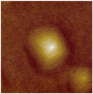
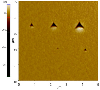
Magnetic Force Microscopy (MFM)
MFM mode with Park AFM measures the magnetic variations over a sample surface by detecting the interaction between a magnetized cantilever and the sample surface. The cantilever measures surface topography on the first scan, then lifts and follows either the stored surface topography (lift mode, available only in selected countries) maintaining a constant distance from the sample surface or a variable distance above the sample surface at a fixed height.
Specifications :
Lateral resolution: 20 nm (depending on the tip size of the cantilever used)Phase resolution: 0.01°
Tunable Magnetic Field MFM (TM-MFM)
TM-MFM mode with Park AFM measures the magnetic domain distribution with respect to magnetic field change.
Specifications :
Requires a magnetic field generator accessory.Adjustable magnetic field of -300 gauss ~ +300 gauss
Field resolution: 3 gauss
Magnetic Field Generator
The magnetic field generator applies an external magnetic field to the sample. The field can be changed from ±300 gauss and is parallel to the sample surface. The changes in magnetic structure can be observed through one of Park AFM’s MFM options.
Specifications :
Applying external magnetic field parallel to sample surfaceTunable magnetic field
Range: -300 - 300 gauss
Composed of pure iron core & two solenoid cells
Mechanical Properties
Park Systems offers a set of mechanical scanning modes that easily measure the mechanical properties of a sample. Each mode features Park AFM’s trademark accuracy that allows users to collect reliable data.
Force Modulation Microscopy (FMM)
Park AFM’s FMM mode measures mechanical property variations over a sample’s surface. An AC modulation is applied to the cantilever while in contact with the sample surface, allowing you to monitor changes in amplitude and phase and gain insights into qualitative elastic and viscous responses.
Specifications :
Adjustable modulation frequency: 1 kHz - 600 kHzFrequency resolution: 0.02 Hz
Amplitude detection: < 0.1 nm
Phase resolution: 0.005°
Nanoindentation
Park AFM’s Nanoindentation mode uses excessive force on the cantilever to indent the sample and measure its mechanical properties including hardness and elasticity.
Specifications :
Batch measurement on user specified points or gridIndenting travel range: 12 µm
Displacement resolution: 0.1nm
Load Application: Piezoelectric Actuator
Maximum Load: 100 nN ~ 100 µN
Load Resolution: 100 nN
Nanolithography
Park AFM’s Nanolithography mode allows users to manipulate and create patterning on the sample surface through applied force or voltage. Tip position for lithography can be easily controlled by importing customized vector drawings or raster (bitmap) images.
Specifications :
Lithography method: vector and/or raster scanBias range: ±10V
Bias noise: 20 μV
High Voltage Toolkit required for voltages over ±10V
Thermal Properties
Park Systems offers a scanning thermal mode that easily measures the thermal properties of a sample. This mode features Park AFM’s trademark accuracy that allows users to collect reliable data.
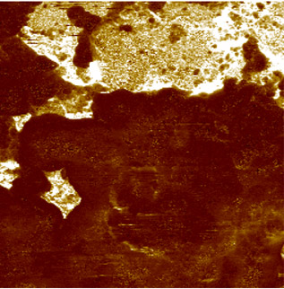
Scanning Thermal Microscopy (SThM)
The SThM mode of Park AFM allows the user to find the local thermal conductivity of a sample by measuring heat transfer between tip and sample using a micro-fabricated probe.
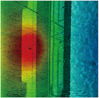
Temperature map of the Pole-Tip-Recession of an active hard disk slider Scan size 20µm x 20µm
Specifications :
Operational modes: Conductivity Contrast Microscopy, Thermal Contrast MicroscopyTip radius and device material of the probe: 100 nm, NiCr & Pd
Thermal spatial resolution: < 100 nm
Allowable current limit: 2.16 mA
Operating temperature: up to 150 °C
Temperature resolution: 0.1 °C
Noise level in temperature: 0.05 °C