Imaging Modes
True Non-Contact™ Mode
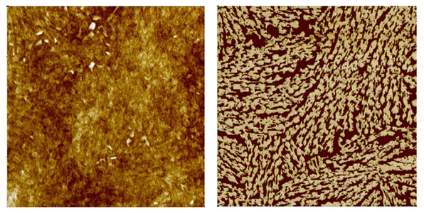
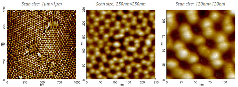
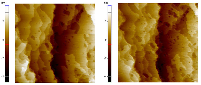
Consistently produces high resolution and accurate data while maintaining the integrity of sample.
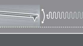
The True Non-Contact mode preserves tip sharpness and sample surface, and you can get more accurate results. In the True Non-Contact mode, a piezoelectric modulator vibrates a cantilever at small amplitude and a fixed frequency near the resonant frequency of the cantilever. As the tip is brought closer to the sample, the van der Waals attractive force between tip and sample changes the amplitude and the phase of the cantilever’s vibration. These changes are monitored by the patented Z-servo feedback system of the Park AFMs, which maintains a tip-surface distance of just a few nanometers without damaging the sample surface or the tip end.
Read More
Contact Mode
The simplest scanning method to image the surface
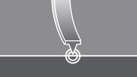
The contact mode is the simplest way to acquire the sample topography. The topography signal comes from the Z scanner position, which maintains the deflection of the cantilever constant on the sample surface.
Read More
Tapping Mode
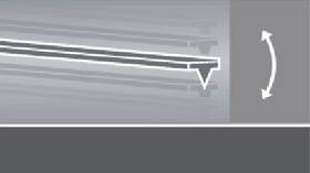
In this alternative technique to non-contact mode, the cantilever again oscillates just above the surface, but at a much higher amplitude of oscillation. The bigger oscillation makes the deflection signal large enough for the control circuit, and hence an easier control for topography feedback. It produces modest AFM results but blunts the tip’s sharpness at a higher rate, ultimately speeding up the loss of its imaging resolution.
Read More