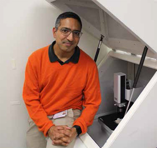
Manish Butte explains the critical role of atomic force microscopes in conducting T-cell research at stanford school of medicine.
Professor Manish Butte works at stanford school of medicine where he teaches a course in immunology and conducts research in the laboratory seeking out the answers to some of the most perplexing questions about the interaction of cells and our immune systems. The laboratory's goal is to address fundamental and therapeutic questions in immunology using innovative nanotechnological and biophysical approaches to visualize and manipulate cells. The primary focus is on understanding the molecular controls that balance T-cell activation versus tolerance. The ultimate aim of our work is to manipulate T-cell signaling pathways to control immunologically-mediated diseases.
Manish J Butte has been awarded 5 Patents including a the 2010 Patent awarded for work with AFM and live Cells: Manish J Butte, Marc Amor Bruce, Jianwei Liu. "United States Patent US 13/307,882 Atomic force microscope manipulation of living cells", The Board Of Trustees Of The Leland Stanford Junior University, Nov 30, 2010
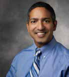
Manish J. Butte, MD PhD is an Assistant Professor in the Department of Pediatrics at Stanford University. He studied Physics at Brown University where he earned his Sc.B. with honors in 1993, studying mathematical neural networks in Prof. Leon Cooper’s group. Afterwards, he earned his M.D. degree from the Brown University School of Medicine in 1996. He then studied protein crystallography under Prof. Robert Fletterick at UCSF and graduated with a Ph.D. in Biophysics in 2000. Returning to clinical training, he completed a Pediatrics residency at the Children's Hospital of Philadelphia in 2003 and a clinical fellowship in Allergy & Immunology at Boston Children's Hospital in 2006, where he specialized in the care of children with immunodeficiencies, autoimmunity, auto-inflammatory disorders, asthma, and allergies. He is board certified in both Pediatrics and Allergy & Immunology. During a joint post-doctoral fellowship at Harvard Medical School (under Prof. Arlene Sharpe) and in the Harvard Chemistry & Chemical Biology Dept. (under Prof. George Whitesides), he worked on T-cell inhibitory pathways and development of microfabricated tools to capture and study immune cells. He transitioned to Stanford in 2009 to start his own lab. His group addresses fundamental, long-standing questions in immunology using innovative nanotechnological approaches to visualize and manipulate cells. His group has pioneered use of biological atomic force microscopy to study immune cell function in health and disease. He cares for children and adults with immunological diseases at the Lucile Packard Children’s Hospital at Stanford and at Stanford Hospital & Clinics.
How does the identification of T Cells help in research?
T-cells are the master organizers of the immune system. They help fight infections by coordinating the adaptive immune response. They demonstrate "memory" by remembering past infections and fighting more quickly the next time; this memory effect is the basis of vaccines. In organ transplants, T-cells are the main cause of chronic rejection and are the targets of immunosuppressive drugs. In cancer, activation of T-cells is necessary to permanently eliminate tumors. In allergies, T-cells demonstrate a defect in tolerance and become unusually sensitive to foreign antigens that ordinarily would be ignored. In aging, the triggering of T-cells become difficult and infections become harder to fight. The more we learn about T-cells, the better we can diagnose and treat these conditions and diseases.
How does the use of AFM technology improve your ability to work with T-Cells?
Atomic force microscopy has been an important tool for innovation in my lab. We are pushing our study of the immune system into an area of cell biology called mechanobiology. This relatively new field entails studying the sensation and generation of mechanical forces by cells. Using AFM, we have shown that T-cells push and pull on their cellular partners, which turns out to be essential at the molecular level for their activation. Furthermore, we have identified mechanical changes in the cytoskeleton of T-cells, the protein framework that enables cells to rapidly crawl and surveil the body for foreign antigens. These mechanical changes influence how T-cell sense the antigens that are presented to them.
Can you describe how you use AFM to visualize and manipulate cells?
We use AFM in a number of ways. First and foremost, we make tiny, brief indentations in the cells, and by measuring the cantilever's path during the indentation, we can calculate the stiffness of the cytoskeleton at that local portion of the cell. By repeating this measurement over and over at different parts of the cell, we can make a "stiffness map" and track changes in the cytoskeleton. By studying stiffness maps after treating cells with antigens or immunomodulatory drugs, we can study the effects of these perturbations on the mechanical state of the cytoskeleton. The second AFM technique we employ, which I believe we invented ourselves, entails putting molecules that can trigger cells on the cantilever tip. For example, we published putting antigenic proteins on the tip of the cantilever that could trigger mast cells. By touching down onto live T-cells, ligating their receptors with the cantilever-bound ligands and simultaneously imaging the T-cells with an optical microscope, we can learn about how molecular signals are interpreted by T-cells. These biochemical signals can be delivered while the AFM is delivering minute mechanical forces to the cell, and moreover, the AFM can measure the mechanical responses of cells undergoing receptor triggering. These studies are critical for us to understand the fundamental processes of activation and tolerance. This might come as a surprise to some users of AFM, but we almost never do "topography"!
Does using AFM for T Cells and other research help improve the ability to cure and/or treat diseases? How has it improved?
AFM has helped us identify a cytoskeletal pathway in T-cells that promotes activation. We are seeking funding for the next level of research, to test whether this pathway can accelerate vaccines or amplify immune responses during infections.
What other applications do you use AFM for in your research?
We have measured the mechanical properties of biological hydrogels that are used for research in cancer, cardiology, and stem cells. We have measured nanoparticles that will be used for gene therapy. We measured the beating of heart cells that allows an improved understanding of pediatric, inherited cardiac diseases. My group is also are quite active at Stanford in developing and publishing new AFM techniques.
What are features of the Park Systems AFM equipment that help most in your research?
The Park NX10 instrument has allowed us to innovate in improving AFM techniques. Specifically, in collaboration with Prof. Olav Solgaard's group at Stanford, we have been developing a new form of high-bandwidth AFM cantilever that can speed up the mechanical measurements mentioned earlier by over 1000-fold! The NX10 has an open head design that allows us to insert miniature mirrors into the light path, which is key to the sensor's mechanism.
How do you see the field of cell research changing in the future with the use of more advanced imaging equipment?
Super-resolution microscopy is revolutionizing optical microscopy in revealing ever finer, nearly-molecular-sized aspects of cells. AFM still has an order-of-magnitude increased resolution over super-resolution microscopy. With the capability to sense chemicals and surface molecules, AFM will be used to improve our understanding the organization of the cell surface. As mechanobiology pervades fields of cell biology, including cancer, stem cells, and others, AFM will become more and more utilized to measure and influence the mechanical state of cells.
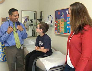
What type of research will you be doing in pediatrics specifically? Will your research help identify the causes?
Currently, our AFM studies of the immune system are focused on fundamentals, but our questions are motivated by the disorders of the children I care for in my clinic with genetic (primary) immune deficiencies. We are inspired to better understand their diseases to improve diagnosis and treatment.