Brian Choi, Bio-application scientist
For more information, please contact app@parksystems.com
Sample courtesy: Dr. Hyuk-soon Choi, Korea Univ. Hospitial
SICM can be used not only in cultured cell imaging but also in biological tissue studies. Tissue-level studies provide different insights compared to cultured cell studies. The naturally organized cells in tissue form a specialized structure for its dedicated function. The alteration of tissue structure can be an indication of losing its function and presence of disease. The phenotype of certain diseases at the tissue level with low resolution has been recorded for pathological purposes. Scanning electron microscopy (SEM) is frequently used as a tissue imaging tool due to wide - range observability and high- resolution imaging capability. However SEM cannot observe biological tissue in aqueous conditions, resulting in skepticism as to its efficacy in physiological studies.
- Observe the naturally organized cells in tissue structure for its dedicated physiological functions.
- Investigate the detail structure of human colon tissue by directly imaging tissue surface morphology in aqueous condition with SICM
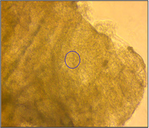 Inverted Optical View
In this study, the morphology of human colon tissue in physiological conditions was visualized with SICM. Normal colon tissue taken from a human large intestine was used for the study. The tissue was soaked in pronase for two hours to remove mucus and fixed with 10% formalin before SICM investigation. The tissue was kept in 1 x PBS (phosphate buffer saline). Since the thickness of colon tissue is about a few hundred micrometers, as shown in the left image, it is not possible to observe surface morphology with an inverted optical microscope. However, SICM successfully imaged colon surface morphology without a complicated sample preparation process. The colon surface was imaged in various scan sizes, from 20 micrometers to five micrometers.
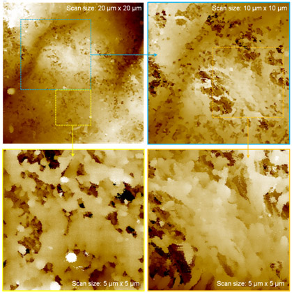 SICM, Human Colon Tissue
As revealed by the specific structure of human colon tissue in the 20-micrometer scan image, the morphology of organized single cells in human colon tissue was clearly observed in the five-micrometer scanned image. Mucus was removed from colon tissue surfaces with pronase treatment. SICM’s physiological tissue imaging offers a new possibility for understanding its functions in detail and relating those functions to structure. It also opens up a new way to investigate the real structural functioning patterns in various tissues. This result indicates that SICM imaging could be developed to provide diagnostic evidence for cancer tissue in the future.
Park Cell Analysis Systems
|
|
|
|
| Park NX12-Bio | Park NX10 | Park XE7 | |
| Scanning Ion Conductance Microscopy (SICM) | |||
| Atomic Force Microscopy (AFM) with liquid probe hand | |||
| Inverted Optical Microscopy (IOM) | |||
| Live Cell Chamber |