Brian Choi, Bio-application scientist
For more information, please contact app@parksystems.com
Cell biologists most frequently use optical cell images, especially for transparent and colourless biological specimens such as cells. In this situation, optical imaging with an inverted optical microscope (IOM) is useful and convenient for determining cell shape and locating edges. However, the optical cell image offers only two-dimensional information on the cell. Although the image seems to offer detailed information about the cell’s inner/outer structures, cells are three-dimensional objects, and this method offers no means of estimating the height information of the cell on the Z axis.
- Distinguishing a cell’s real topography is not possible with inverted optical microscopy (IOM)
- Acquire 3-D cell structure
- Analyze the cell’s volume, size (height/depth), and high-resolution surface image
Technically, the two-dimensional flat cell image is the collection of the applied lights that have passed through the transparent cell. Depending on the refractive cell components in a certain thickness of cell, the angle of the light path changes. The collection of the angle differences is constructed as a two-dimensional cell image. The optical image of the cell displays a mixture of the light signals from all components of the inner/outer cell, making it impossible to distinguish details. Unlike optical microscopic imaging, SICM visualizes cell surface morphology only, generating three-dimensional topography by recording the height information of the cell membrane in a scan area.
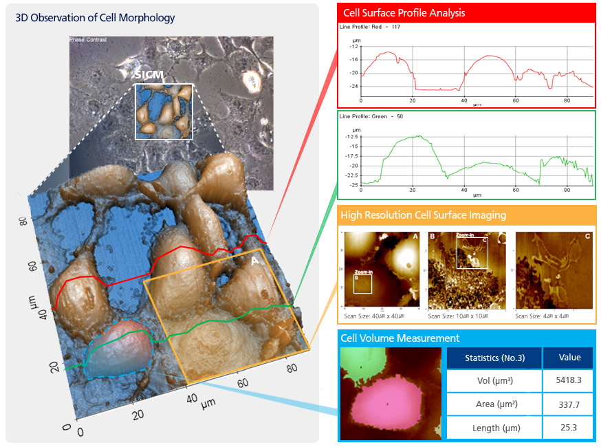
Based on three-dimensional topography data, a researcher can clearly analyze structural and physical properties, such as height, width, depth, length, and volume, in a single cell or multiple cells. Sub-cellular features can also be accurately measured.
Park Cell Analysis Systems
|
|
|
|
| Park NX12-Bio | Park NX10 | Park XE7 | |
| Scanning Ion Conductance Microscopy (SICM) | |||
| Atomic Force Microscopy (AFM) with liquid probe hand | |||
| Inverted Optical Microscopy (IOM) | |||
| Live Cell Chamber |