Optical Properties – SERS/NSOM/Topography
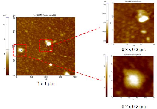
Gold nanoparticles are imaged with high resolution. Gold nanoparticles are known to promote surface-enhanced Raman spectroscopy (SERS). In Ref. 1 below, the authors use this property to demonstrate an ultrasensitive analytical method by exploiting a Raman probe covalently bound to gold nanoparticles; the biological marker to be detected was adsorbed on functionalized gold nanoparticles. The technique resulted in the ability to detect marker concentrations down to the picomolar (pM) level.
Relevant Publications using the XE-series AFM and SERS/NSOM
1. Anna Rita Bizzarri, Salvatore Cannistraro; Surface-enhanced Raman spectroscopy combined with atomic force microscopy for ultrasensitive detection of thrombin; Analytical Biochemistry 393 (2009) 149–154
Equipment: Park Systems XE-100
2. Nanfang Yu, Ertugrul Cubukcu, Laurent Diehl, Mikhail A. Belkin, Kenneth B. Crozier, Federico Capasso, David Bour, Scott Corzine, and Gloria Höfler; Plasmonic quantum cascade laser antenna; APPLIED PHYSICS LETTERS 91, 173113 (2007)
Equipment: Park Systems XE-120
3. Sangwook Oh, Chankyeong Hyon, Sanghoon Sull, Sungwoo Hwangb, Yongju Park; Detection and volume estimation of semiconductor quantum dots from atomic force microscope images; Rev. Sci. Instrum., Vol. 74, No. 11, (2003) 4687 - 4695
Equipment: Park Systems XE-100
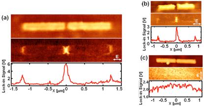
Fig 4. (color online) Mid-ir apertureless-NSOM imaging of plasmonic laser antennas. (a) Top and middle panels: simultaneous AFM topography and NSOM image for a resonant antenna with L=12 μm fabricated on the facet of a λ=7.0 μm QCL. Bottom panel: corresponding line scan along the antenna axis. (b) Top and middle panels: simulaneous AFM topography and NSOM image for a resonant antenna with L=0.76 μm fabricated on the facet of a λ=5.3 μm QCL. Bottom panel: corresponding line scan along the antenna axis. (c) Top and middle panels: simultaneous AFM topography and NSOM image of an antenna with L=0.79 μm fabricated on the facet of a λ=5.3 μm QCL but with orientation normal to the laser field. Bottom panel: corresponding line scan along the antenna axis. The polarization of the incident electric field is indicated in each NSOM image.